


In total anomalous pulmonary venous return, the pulmonary veins, blood vessels that normally carry oxygenated blood from the lungs to the left side of the heart, connect instead to the right side of the heart. This blood then flows to the left heart through a hole in the wall separating the left and right heart chambers.
Children with more mild forms of TAPVR may only have mild feeding and breathing problems in infancy.
Children with more severe TAPVR may have bluish discoloration of the skin (cyanosis), shortness of breath, and fatigue.
Children with the most severe forms of this defect may develop severe respiratory distress and cyanosis shortly after birth.
Echocardiography is needed for diagnosis.
The defect is treated with surgical repair.
(See also Overview of Heart Defects.)
Total anomalous pulmonary venous return (TAPVR) accounts for 1 to 2% of birth defects of the heart.
Normally, the pulmonary veins return blood that has picked up oxygen in the lungs to the left atrium. This oxygenated blood then flows from the left atrium into the left ventricle, where it is pumped to the body. (See also Normal Fetal Circulation.) In infants born with TAPVR, the pulmonary veins do not connect normally to the left atrium and connect instead by an abnormal pathway eventually leading to the right atrium. So the right atrium, which usually receives only deoxygenated blood from the body to be pumped to the lungs, receives a mix of oxygenated and deoxygenated blood. The additional blood return causes an increased workload and enlargement of the right side of the heart. Even more importantly, blood flow from the pulmonary veins makes its way to the right atrium through various pathways, going up above the heart, down below the heart, or around the back wall of the heart. These pathways can be narrow or blocked, causing blood to back up in the lungs and pressure to build up in the lungs, preventing their normal function. Blood only flows to the left side of the heart through a hole between the right and left atria (a patent foramen ovale or atrial septal defect).
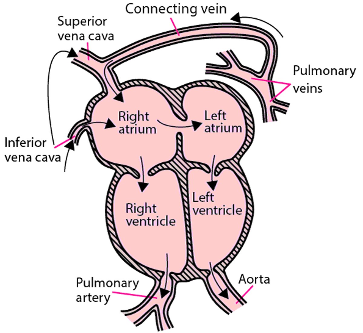
Total Anomalous Pulmonary Venous Return
The pulmonary veins do not connect to the left atrium; instead, the entire pulmonary venous return enters systemic venous circulation through various connections (in this case the connecting vein is supracardiac or above the heart). Connecting veins can also be below the heart (infracardiac) or behind the heart. Systemic blood flow depends on right-to-left atrial shunting. AO = aorta; IVC = inferior vena cava; LA =left atrium; LV = left ventricle; PA = pulmonary artery; PV =pulmonary veins; RA = right atrium; RV = right ventricle; SVC = superior vena cava. |

Symptoms of TAPVR
Newborns with a severe form of TAPVR have difficulty breathing and bluish coloration of the skin (cyanosis).
In milder forms, symptoms of heart failure (see figure Types of Heart Failure) may be present but be more difficult to detect. Symptoms of heart failure include shortness of breath, a bluish color to the skin (cyanosis), and fatigue. The level of oxygen is usually lower than normal, but some infants may have no symptoms or cyanosis.
Diagnosis of TAPVR
Echocardiography
Doctors suspect the diagnosis based on the findings on a chest x-ray, a heart murmur heard with a stethoscope, or low oxygen level in the blood detected by pulse oximetry. The diagnosis is confirmed by echocardiography (ultrasonography of the heart). Occasionally, magnetic resonance imaging (MRI) of the heart or computed tomography (CT) angiography may be needed so doctors can more clearly view the defect.
Treatment of TAPVR
Surgical repair
Medication to treat heart failure before surgery
Newborns with TAPVR require surgery early in life. When there is severe obstruction in the pulmonary venous pathway, emergency surgical repair is often needed. Heart failure should be treated with drugs to improve breathing until surgery can be done.
Surgical repair consists of creating a connection between the pulmonary veins and the left atrium. The abnormal connection pathway from the left atrium is tied off.
Some children need to take antibiotics before visits to the dentist and before certain surgeries (such as on the respiratory tract). These antibiotics are used to prevent a serious heart infection called endocarditis.
More Information
The following English-language resources may be useful. Please note that THE MANUAL is not responsible for the content of these resources.
American Heart Association: Common Heart Defects: Provides an overview of common birth defects of the heart for parents and caregivers
American Heart Association: Infective Endocarditis: Provides an overview of infective endocarditis, including summarizing antibiotic use, for parents and caregivers


