


Birth defects are more common in the kidney and urinary system (urinary tract) than in any other system of the body. Defects can develop in the
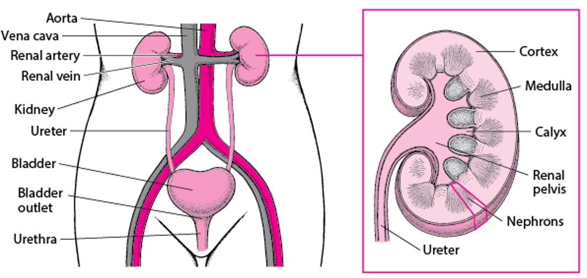
Kidneys—the two organs that filter waste from the blood to make urine
Ureters—the tubes that transport urine from the kidneys to the bladder
Bladder—the expandable, muscular sac that holds urine
Urethra—the tube that drains urine from the bladder out of the body
Each kidney continuously produces urine, which then drains through the ureter into the bladder at a low pressure. From the bladder, urine drains through the urethra to exit the body. In males, the urethra is located in the penis. In females, the urethra ends in the vulvar area (the area of the external female genital organs). Usually, urine is free of bacteria and other infectious organisms.
A Look Inside the Urinary Tract

Did You Know...
|
Complications of urinary tract defects
Urinary tract defects may
Block or slow the flow of urine
Allow urine to flow backwards from the bladder to the kidneys (urinary reflux)
Any birth defect that blocks or slows the flow of urine can cause urine to become stagnant, which can result in urinary tract infections (UTIs) or formation of kidney stones. If the flow of urine is blocked, it can cause pain or damage to the kidney.
Urinary reflux usually happens when defects involve the junction where a ureter connects to the bladder. Normally the junction allows urine to flow only one way, from the kidneys to the bladder. Defects of the junction can allow urine to flow backward from the bladder into the kidney (urinary reflux). In addition, other defects that block the flow of urine can increase the pressure in the bladder and cause urinary reflux. Reflux can affect one side or both sides.
Urinary reflux and/or frequent infections can damage the kidneys and ureters over time. Kidney damage can cause high blood pressure and rarely kidney failure.
Severe urinary tract defects in a fetus can cause little or no urine to be produced. The fetus's urine becomes part of the fluid that surrounds the fetus in the uterus (called amniotic fluid). If the fetus does not release enough urine, the amount of amniotic fluid is reduced. If there is too little amniotic fluid, the fetus's lungs, heart, face, and limbs may develop abnormally. Severe defects can cause death while the fetus is in the womb or shortly after birth.
Symptoms of Kidney and Urinary Tract Birth Defects
Many urinary tract defects cause no symptoms and are often discovered only when imaging studies are done for other reasons, or during a well-child examination. Some kidney defects do not cause problems or become known until adulthood.
When urinary tract defects do cause symptoms, children may have
Blood in their urine (hematuria)
Recurring symptoms of urinary tract infections
Recurring involuntary release of urine (urinary incontinence)
Abdominal pain, and/or vomiting due to blockage (obstruction) of urine flow
Children who have urinary obstruction are also at increased risk for significant urinary bleeding after a minor injury because the kidney is under pressure.
Diagnosis of Kidney and Urinary Tract Birth Defects
Before birth, prenatal ultrasonography and blood tests
After birth, imaging tests and sometimes cystoscopy
Before birth, urinary tract defects are often discovered by doctors during routine prenatal ultrasonography or other routine screening tests for hereditary disorders.
After birth, if doctors suspect a child has a urinary defect, they typically do imaging tests such as ultrasonography, computed tomography (CT), nuclear scans, and magnetic resonance imaging (MRI). Sometimes, doctors do intravenous urography or cystoscopy. In cystoscopy, doctors look inside the bladder and urethra through a flexible viewing tube called a cystoscope (a type of endoscope).
To diagnose certain defects of the urinary tract, doctors sometimes do a test called voiding cystourethrography (VCUG). For voiding cystourethrography, a catheter is passed through the urethra into the bladder, a liquid that shows up on x-rays (contrast agent) is put through the catheter, and x-rays are taken before, during, and after the child urinates.
As children grow, these tests may be repeated at scheduled intervals to assess how the bladder, ureters, urethra, and kidneys are developing or functioning and to assess if the child has outgrown the defect.
Treatment of Kidney and Urinary Tract Birth Defects
Sometimes surgery
Defects that cause symptoms or those that lead to increased pressure in the kidneys or bladder usually need to be surgically corrected.


