


Hypophosphatemic rickets is a disorder in which the bones become painfully soft and bend easily because the blood contains low levels of the electrolyte phosphate.
(See also Introduction to Congenital Kidney Tubular Disorders.)
This very rare disorder is nearly always hereditary. It is most commonly inherited as a dominant gene that is carried on the X chromosome, one of the two sex chromosomes. Because only one gene is needed when a dominant gene is involved, one of the parents is likely to have the syndrome. Siblings of children with the disorder have a 50% chance of having it. There are several other even rarer forms of hypophosphatemic rickets.
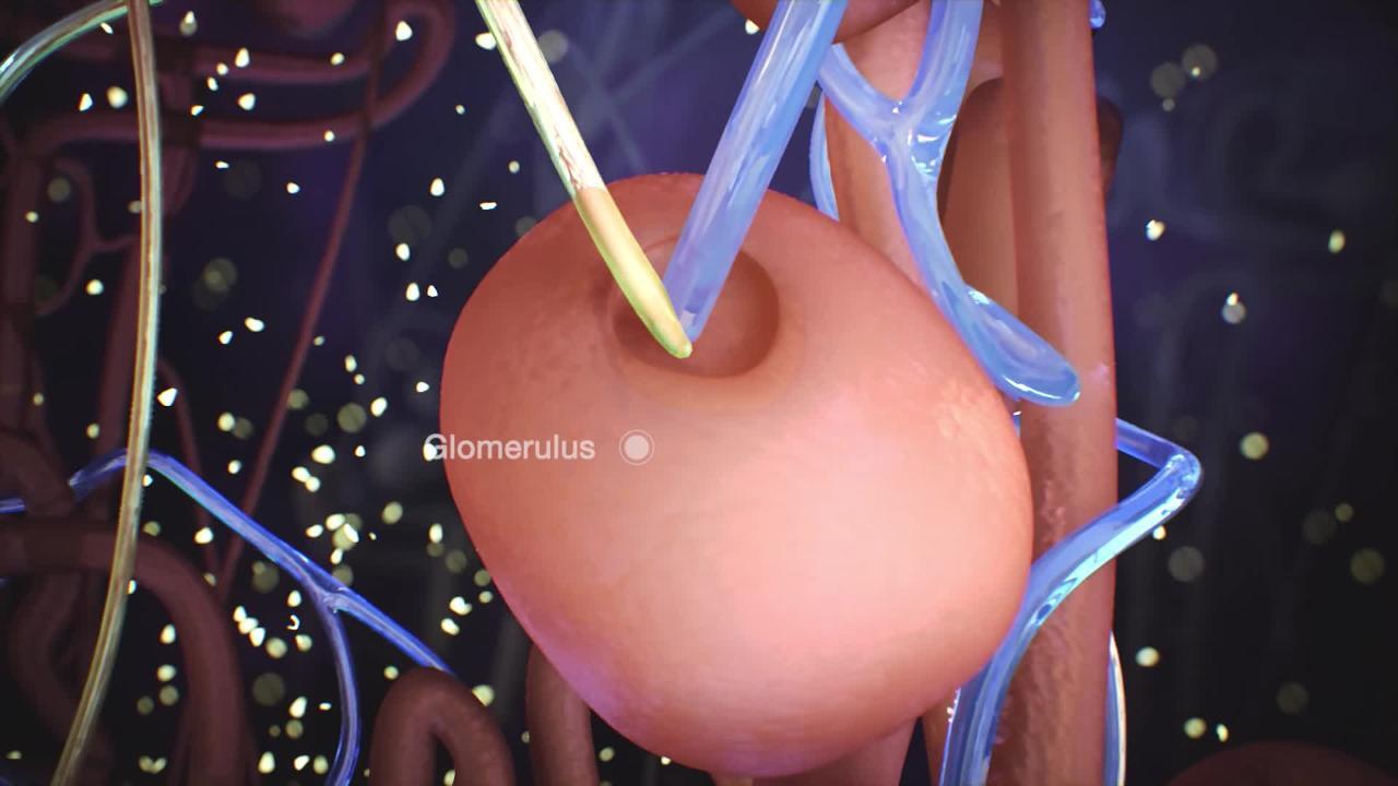
The genetic defect causes a kidney tubule abnormality that allows an inappropriately high amount of phosphate to be excreted into the urine, resulting in low levels of phosphate in the blood. Because bones need phosphate for growth and strength, this deficiency causes defective bones.
In rare cases, the disorder develops as a result of certain cancers, such as giant cell tumors of bone, sarcomas, prostate cancer, and breast cancer. Hypophosphatemic rickets is not the same as rickets caused by .
Symptoms of Hypophosphatemic Rickets
Hypophosphatemic rickets usually begins to cause abnormalities in the first year of life. Abnormalities may be so mild that they cause no noticeable symptoms or so severe that they cause bowing of the legs and other bone deformities, bone pain, joint pain, and poor bone growth with short stature. Bony outgrowths where muscles attach to bones may limit movement at those joints.
The space between a baby’s skull bones may close too soon (called craniosynostosis). If the skull bones close too soon, the child's growing brain has no room to expand and pressure increases within the brain. The increased pressure on the brain can cause developmental abnormalities.
Diagnosis of Hypophosphatemic Rickets
Blood and urine tests
Sometimes bone x-rays
Sometimes genetic testing
Laboratory tests show that calcium levels in the blood are normal, but phosphate levels are low.
Urine is also tested to detect the levels of phosphate that have been excreted. The phosphate levels in the urine are high.
Doctors may also take x-rays of bones.
Genetic testing can help confirm the diagnosis.
Siblings of children who are affected should have a medical evaluation, including laboratory testing, imaging tests, and sometimes genetic testing. Genetic testing also may be offered to other family members.
Treatment of Hypophosphatemic Rickets
Phosphate and calcitriol
Burosumab for the most common form of hypophosphatemic rickets
Sometimes surgical removal of tumors
Treatment of hypophosphatemic rickets is aimed at raising phosphate levels in the blood, which promotes normal bone formation.
calcitriol must be adjusted carefully because this treatment often leads to high levels of calcium in the blood and urine, a build up of calcium in the kidneys, or kidney stones. These side effects can harm the kidneys and other tissues.
People who have the most common form of hypophosphatemic rickets are given the drug burosumab and are not given the phosphate and calcitriolmonoclonal antibody and is given as an injection under the skin. It helps increase the levels of phosphorus in the blood, helps decrease the severity of rickets, and in children may increase height. Burosumab can be given to children and adults.


