


Pulmonary function tests measure the lungs' capacity to hold air, to move air in and out, and to absorb oxygen.
Pulmonary function tests are better at detecting the general type and severity of lung disorder than at defining the specific cause of problems; however, these tests can be used to diagnose some specific disorders, such as asthma and chronic obstructive pulmonary disease (COPD).
(See also Medical History and Physical Examination for Lung Disorders and Overview of the Respiratory System.)
Lung flow rate measurements
The assessment of a lung disorder often involves testing
How much air the lungs can hold (lung volume)
How much and how quickly air can be exhaled (airflow)
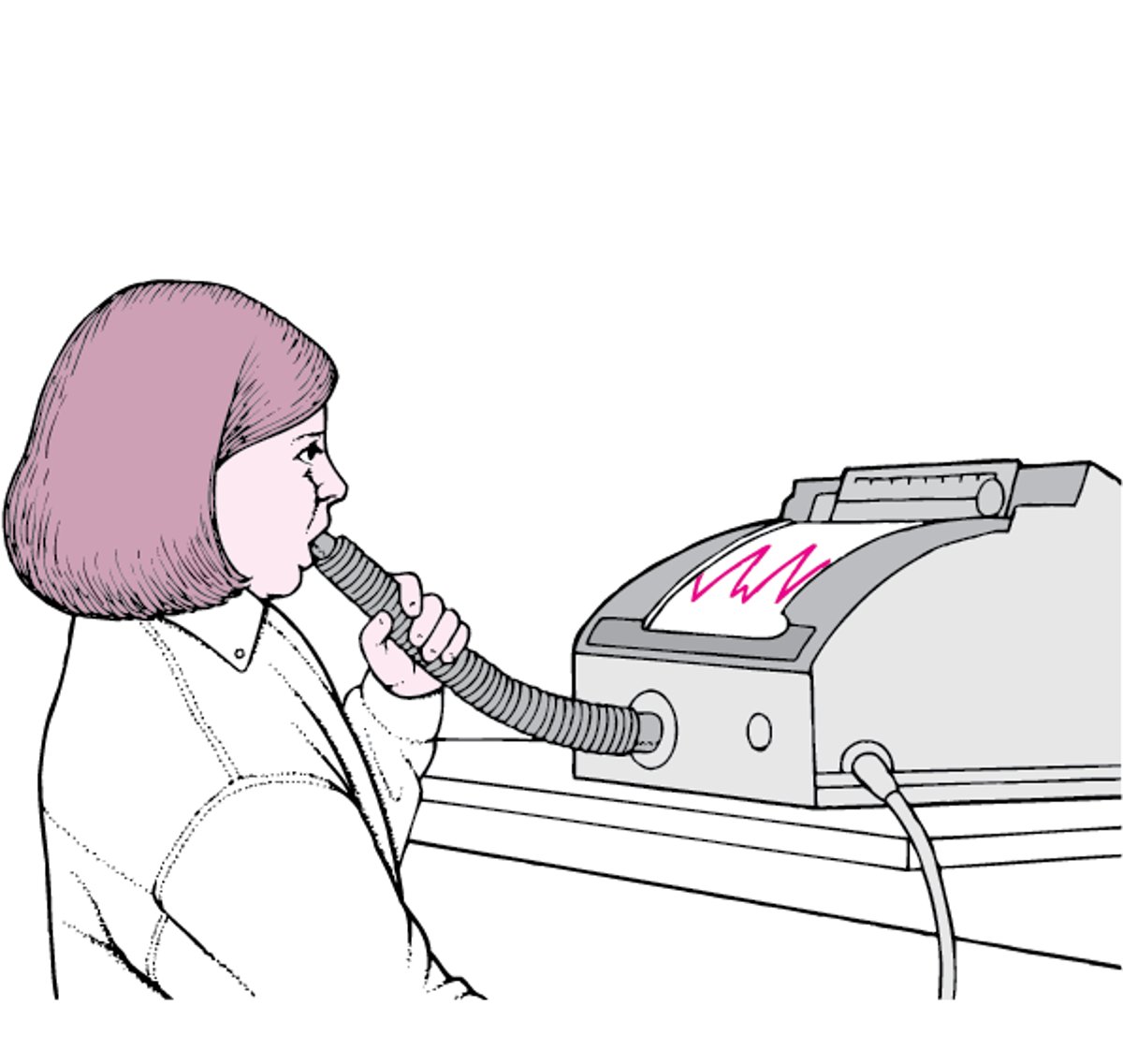
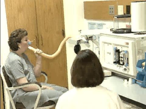
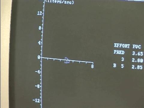
Airflow measurements are made with a spirometer, which consists of a mouthpiece and tubing connected to a recording device. The person’s lips should be held tightly around the mouthpiece, and nose clips should be worn to ensure that all the air inhaled or exhaled goes through the mouth. A person inhales deeply, then exhales forcefully as quickly as possible through the tubing while measurements are taken. The volume of air inhaled and exhaled and the length of time each breath takes are recorded and analyzed. This measurement is repeated several times to be sure the results are consistent. Often, the tests are repeated after a person takes a medication that opens the airways of the lungs (bronchodilator).
In disorders such as asthma and chronic obstructive pulmonary disease (COPD), the ability to exhale quickly is impaired.
Using a Spirometer
A spirometer consists of a mouthpiece, tubing, and a recording device. To use a spirometer, a person inhales deeply, then exhales vigorously and as quickly as possible through the tubing. The recording device measures the volume of air inhaled or exhaled and the length of time each breath takes. |

Video from Lahey Clinic Media Center.
Video from Lahey Clinic Media Center.


Video from Lahey Clinic Media Center.

Video from Lahey Clinic Media Center.
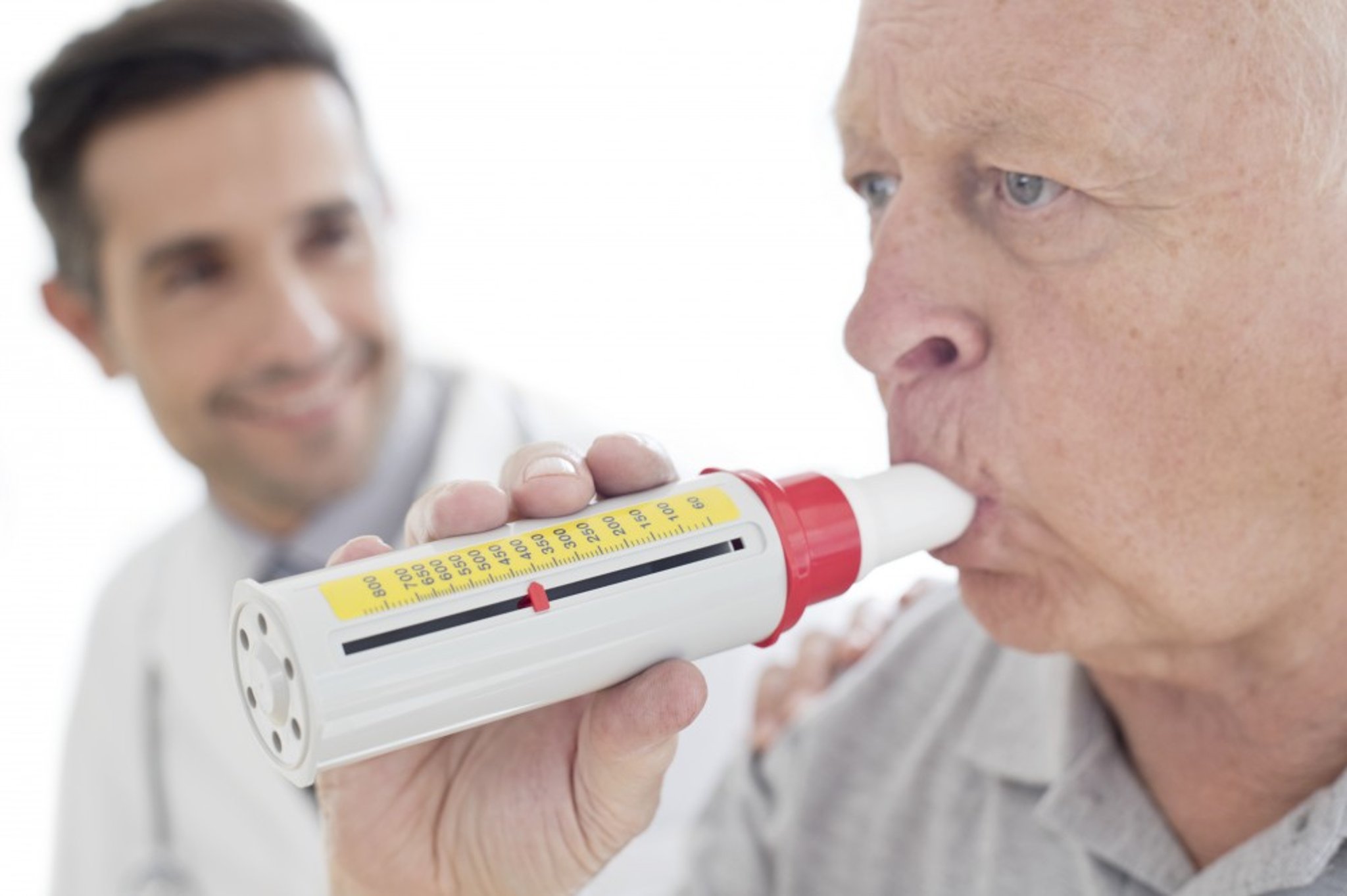
A simpler device for measuring how quickly air can be exhaled is the small, hand-held peak flow meter. After inhaling deeply, a person blows into this device as hard as possible.

SCIENCE PHOTO LIBRARY
Lung volume measurement
Lung volume measurements reflect the stiffness or elasticity of the lungs and rib cage as well as the strength of respiratory muscles. The lungs are abnormally stiff in disorders such as pulmonary fibrosis, and the chest wall is abnormally stiff in disorders such as curvature of the spine (scoliosis). Various neuromuscular disorders such as myasthenia gravis and Guillain-Barré syndrome can cause weakness of the diaphragm and other respiratory muscles, decreasing the volume of air in the lungs. Increased stiffness of the lungs causes lower lung volume measurements. In disorders such as COPD, decreased elasticity of the lungs makes it difficult to breathe out. More air is trapped in the lungs, causing higher than expected lung volume measurements.
Lung volume measurements made using spirometry are only estimates. More accurate measurements can be made using
Body plethysmography
Gas dilution
In body plethysmography, a person sits inside an airtight plastic box. Because the box is airtight, the volume of air the person breathes in and the change in air pressure during breathing can be measured. A computer calculates lung volumes based on these measurements.
Using gas dilution, a person breathes in a known amount of a gas, usually helium. A computer calculates lung volume based on how much gas the person exhales.
Flow rate testing
Most spirometers can continuously display lung volumes and flow rates during a forced breathing maneuver. These flow rates can be particularly helpful in detecting abnormalities that partially block the voice box (larynx) and windpipe (trachea).
Muscle strength assessment
The strength of the respiratory muscles can be measured by having the person forcibly inhale and exhale against a pressure gauge. Disorders that weaken the muscles, such as muscular dystrophy and amyotrophic lateral sclerosis (ALS, or Lou Gehrig disease), weaken respiratory muscles and make breathing more difficult. Muscle strength can also be assessed by having the person do spirometry while sitting up and while lying down.
Diffusing capacity measurement
A diffusing capacity test can estimate how efficiently oxygen is transferred from the air sacs of the lungs (alveoli) to the bloodstream. Because the diffusing capacity of oxygen is difficult to measure directly, a person inhales a small amount of carbon monoxide, holds the breath for 10 seconds, and then exhales into a carbon monoxide detector.
If the test shows that carbon monoxide is not well absorbed, oxygen will not be exchanged normally between the lungs and the bloodstream either. The diffusing capacity is characteristically abnormal in people with pulmonary fibrosis, in those with disorders affecting the blood vessels of the lungs, and in some people with chronic obstructive pulmonary disease (COPD).
Maximal voluntary ventilation (MVV)
MVV measures a person's maximum overall ability to breathe. This test is done in the sitting position. A person is instructed to breathe as rapidly and deeply as possible through a spirometer for a predetermined period of time, usually 15 to 30 seconds. The volume of air moved over that period of time is measured. MVV will be decreased in diseases that affect airflow or weaken respiratory muscles. Because this test is dependent upon the ability of a person to cooperate, it is not used as often as other lung function tests.


