


Infectious mononucleosis is caused by Epstein-Barr virus (EBV, human herpesvirus type 4) and is characterized by fatigue, fever, pharyngitis, and lymphadenopathy. Fatigue may persist weeks or months. Severe complications, including airway obstruction, splenic rupture, and neurologic syndromes, occasionally occur. Diagnosis is clinical or with EBV serologic testing. Treatment is supportive.
EBV is a herpesvirus that infects 50% of children before age 5 (1). Over 90% of adults are seropositive for EBV. Its host is humans.
EBV infection is usually asymptomatic.
(See Overview of Herpesvirus Infections.)
Reference
1. Johannsen EC, Kaye KM: Epstein-Barr Virus (Infectious Mononucleosis, Epstein-Barr Virus–Associated Malignant Diseases, and Other Diseases). In Mandell, Douglas, and Bennett's Principles and Practice of Infectious Diseases (Ninth Edition), Elsevier, 2020, pp. 138, 1872-1890, 2020 . ISBN: 9996119890, 9789996119897
Pathophysiology of Infectious Mononucleosis
After exposure in the oral cavity, EBV infects B lymphocytes. Morphologically abnormal (atypical) lymphocytes develop, mainly from CD8+ T cells that respond to the infection.
After primary infection, EBV remains within the host, primarily in B lymphocytes, for life and undergoes intermittent asymptomatic shedding from the oropharynx. The virus is detectable in oropharyngeal secretions of 10 to 20% of healthy EBV-seropositive adults (1). Shedding increases in frequency and titer in patients who are immunocompromised (eg, organ allograft recipients, people living with HIV).
EBV has not been recovered from environmental sources and is not very contagious.
Transmission
Transmission may occur via transfusion of blood products but much more frequently occurs via kissing between an uninfected and an EBV-seropositive person who is shedding the virus asymptomatically. Only about 5% of patients acquire EBV from someone who has acute infection (1).
Early childhood transmission occurs more frequently among lower socioeconomic groups and in crowded conditions.
Associated disorders
EBV is statistically associated with and likely has a causal role in
Certain B-cell tumors in patients who are immunocompromised
Certain forms of Hodgkin lymphoma
Certain gastric cancers
EBV does not cause chronic fatigue syndrome. However, it rarely causes a syndrome that may include fever, interstitial pneumonitis, pancytopenia, hepatitis, or uveitis (ie, chronic active EBV).
Pathophysiology reference
1. Johannsen EC, Kaye KM: Epstein-Barr Virus (Infectious Mononucleosis, Epstein-Barr Virus–Associated Malignant Diseases, and Other Diseases). In Mandell, Douglas, and Bennett's Principles and Practice of Infectious Diseases (Ninth Edition), Elsevier, 2020, pp. 138, 1872-1890, 2020 . ISBN: 9996119890, 9789996119897
Symptoms and Signs of Infectious Mononucleosis
In most young children, primary EBV infection is asymptomatic. Symptoms of infectious mononucleosis develop most often in older children and adults.
The incubation period is about 30 to 50 days. Fatigue can last for months but is usually maximal during the first 2 to 3 weeks.
Most patients have the triad of
Fever
Pharyngitis
Adenopathy
Fever usually peaks in the afternoon or early evening, with a temperature around 39.5° C, although it may reach 40.5° C.
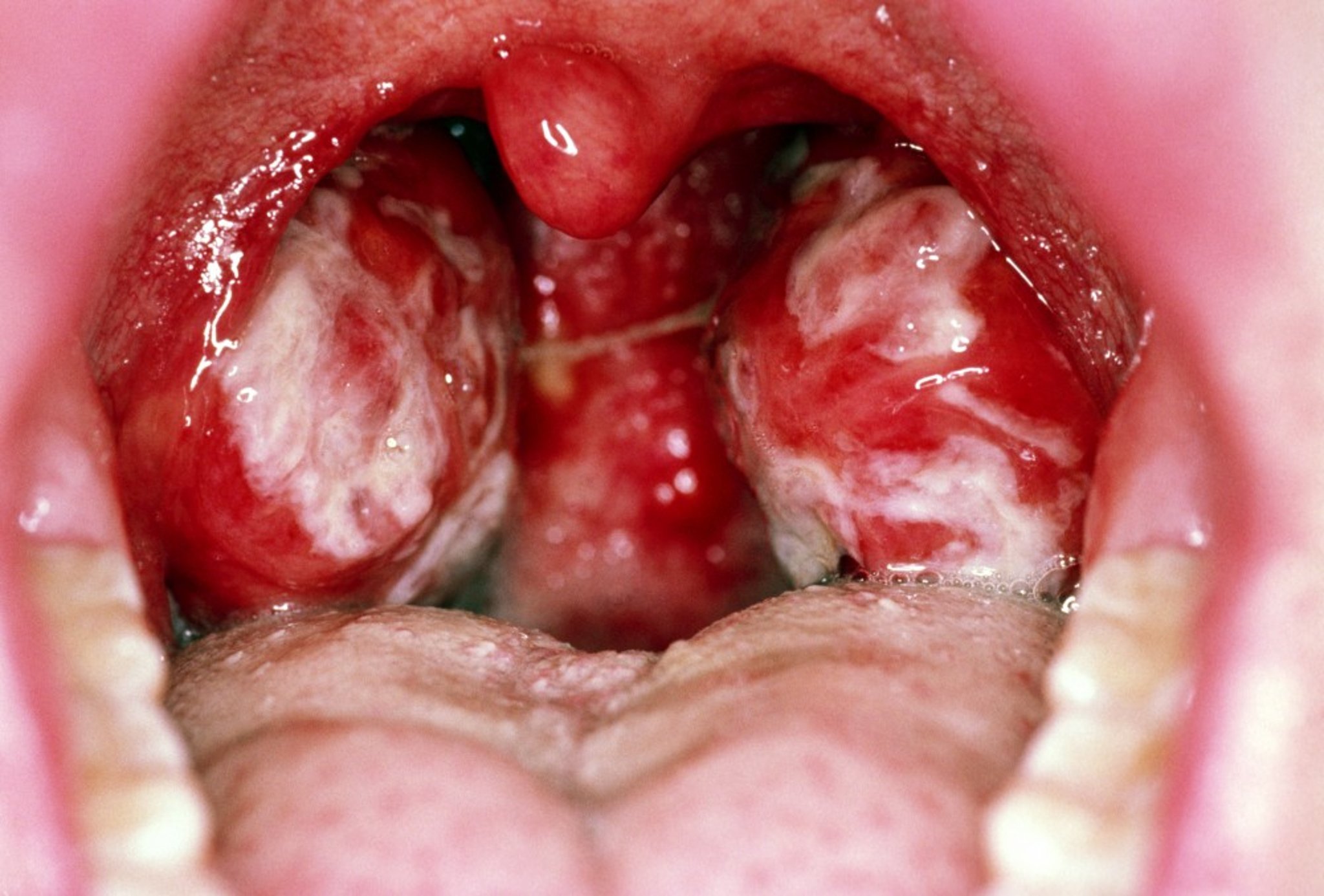
Pharyngitis may be severe, painful, and exudative and may resemble streptococcal pharyngitis.

DR P. MARAZZI/SCIENCE PHOTO LIBRARY


MID ESSEX HOSPITAL SERVICES NHS TRUST/SCIENCE PHOTO LIBRARY
Adenopathy is usually symmetric and may involve any group of nodes, particularly the anterior and posterior cervical chains. Adenopathy may be the only manifestation.
Other symptoms and signs include
Splenomegaly
Mild hepatomegaly and hepatic percussion tenderness
Periorbital edema and palatal petechiae
Less frequently maculopapular eruptions
Rarely jaundice
Splenomegaly, which occurs in about 50% of cases (1), is maximal during the 2nd and 3rd week and usually results in only a barely palpable splenic tip.
Complications
Although recovery is usually complete, complications may be dramatic.
Neurologic complications are rare but may include encephalitis, seizures, Guillain-Barré syndrome, peripheral neuropathy, viral meningitis, myelitis, cranial nerve palsies, and psychosis. Encephalitis may manifest with cerebellar dysfunction, or it may be global and rapidly progressive, similar to herpes simplex encephalitis, but is usually self-limited.
Hematologic complications are usually self-limited. They include
Granulocytopenia
Thrombocytopenia
Hemolytic anemia
Transient mild granulocytopenia or thrombocytopenia occurs in about 50% of patients; severe cases with bacterial infection or bleeding occur less frequently. Hemolytic anemia is often due to anti-i-specific cold-agglutinin antibodies.
Splenic rupture can have severe consequences. It can result from splenic enlargement and capsular swelling, which are maximal 10 to 21 days after presentation. A history of trauma is present only about half of the time. Rupture is usually painful but occasionally causes painless hypotension. For treatment, see Splenic Injury.
Respiratory complications include, rarely, upper airway obstruction due to pharyngeal or paratracheal lymphadenopathy; respiratory complications may respond rapidly to corticosteroids.
Hepatic complications include elevated aminotransferase levels (about 2 to 3 times normal, returning to baseline over 3 to 4 weeks); they occur in about 90% of patients (1). If jaundice or more severe enzyme elevations occur, other causes of hepatitis should be investigated.
Overwhelming infection with EBV occurs sporadically but may cluster in families, particularly those with X-linked lymphoproliferative syndrome. Survivors of overwhelming primary EBV infection with lymphoproliferative syndrome are at risk of developing agammaglobulinemia or lymphoma.
Symptoms and signs reference
1. Johannsen EC, Kaye KM: Epstein-Barr Virus (Infectious Mononucleosis, Epstein-Barr Virus–Associated Malignant Diseases, and Other Diseases). In Mandell, Douglas, and Bennett's Principles and Practice of Infectious Diseases (Ninth Edition), Elsevier, 2020, pp. 138, 1872-1890, 2020 . ISBN: 9996119890, 9789996119897
Diagnosis of Infectious Mononucleosis
Heterophile antibody test
Sometimes EBV serologic testing
Infectious mononucleosis should be suspected in patients with typical symptoms and signs. Exudative pharyngitis, anterior cervical lymphadenopathy, and fever may be clinically indistinguishable from those caused by group A beta-hemolytic streptococci. However, posterior cervical or generalized adenopathy or hepatosplenomegaly suggests infectious mononucleosis. Moreover, detection of streptococci in the oropharynx does not exclude infectious mononucleosis.
Differential diagnosis
Primary HIV infection can produce a clinical picture resembling acute EBV infection. If patients have risk factors for HIV infection, the following should be done:
Quantitative HIV RNA viral load in blood
Combination antibody immunoassay and p24 antigen assay
HIV enzyme-linked immunosorbent assay (ELISA)/Western blot is usually negative during the acute infection and thus should not be used alone to diagnose early primary HIV infection. Quantitative HIV RNA and p24 antigen detection are more sensitive for diagnosing acute HIV infection because HIV RNA and p24 antigen are present in blood before HIV antibodies develop.
Pearls & Pitfalls
|
Cytomegalovirus (CMV) may also cause a mononucleosis syndrome, with atypical lymphocytosis as well as hepatosplenomegaly and hepatitis but usually not with severe pharyngitis.
Toxoplasmosis may cause a syndrome similar to infectious mononucleosis with fever and lymphadenopathy but usually not with pharyngitis.
Laboratory tests
Laboratory diagnosis usually involves a complete blood count and EBV serologic testing. Lymphocytes that are morphologically atypical account for up to 30% of the white blood cells. Although individual lymphocytes may resemble leukemic lymphocytes, lymphocytes are heterogeneous, which is unlikely in leukemia. Atypical lymphocytes may also be present in HIV or CMV infection, hepatitis B, influenza B, rubella, or other viral illnesses, so diagnosis requires serologic testing. However, very high atypical lymphocyte counts are typically seen only in primary EBV and CMV infection.
Two serologic tests are used to diagnose acute EBV infection:
Heterophile antibody testing
Specific EBV antibody testing
Heterophile antibodies are measured using various agglutination card (monospot) tests. However, heterophile antibodies are present in only 50% of patients < 5 years and in about 80 to 90% of adolescents and adults with infectious mononucleosis. Importantly, the heterophile antibody test may be false-positive in some patients with acute HIV infection. The titer and prevalence of heterophile antibodies rise during the 2nd and 3rd week of illness. Thus, if the diagnosis is suspected and the heterophile antibody test is negative early in clinical illness (in the first week), testing can be repeated approximately 7 days later. Due to the potential for false positive or negative results, the Centers for Disease Control and Prevention (CDC) does not recommend heterophile antibodies to diagnose primary EBV infection (see CDC: Laboratory Testing). However, a positive heterophile antibody test in the appropriate clinical situation is generally sufficient to confirm the diagnosis of primary EBV. Alternatively, EBV antibody testing can be performed.
EBV-specific antibody testing is highly sensitive. The presence of IgM antibodies to the EBV viral capsid antigen (VCA) indicates primary EBV infection (these antibodies disappear within 3 months after infection). IgG VCA (EBV VCA-IgG) also develops early in primary EBV infection, but these antibodies persist for life. EBV nuclear antigen (EBNA-IgG) antibodies develop later (after 2 to 4 months ) in acute EBV infection and also persist for life. If EBV antibody titers are negative or indicate remote infection (ie, positive for IgG antibodies and negative for IgM antibodies), other diagnoses that can present with similar symptoms (eg, acute HIV infection, CMV infection) should be considered.
Treatment of Infectious Mononucleosis
Supportive care
Corticosteroids possibly helpful for severe disease
Treatment of infectious mononucleosis is supportive. Patients are encouraged to rest during the acute phase but can resume activity when fever, pharyngitis, and malaise abate. To prevent splenic rupture, patients should avoid heavy lifting and contact sports for 1 month after presentation and until splenomegaly (which can be monitored by ultrasonography) resolves.
Prognosis for Infectious Mononucleosis
Infectious mononucleosis is usually self-limited. Duration of illness varies; the acute phase lasts about 2 weeks. Generally, 20% of patients can return to school or work within 1 week, and 50% within 2 weeks. Fatigue may persist for several more weeks or, in up to 10% of cases, for months.
Death occurs in < 1%, mostly resulting from complications (eg, encephalitis, splenic rupture, airway obstruction).
Key Points
EBV infection is very common; the virus remains within the host for life and is intermittently and asymptomatically shed from the oropharynx.
Only about 5% of patients acquire EBV from someone who has acute infection.
Typical manifestations include fatigue (sometimes persisting weeks or months), fever, pharyngitis, splenomegaly, and lymphadenopathy.
Uncommon severe complications include encephalitis and other neurologic manifestations, splenic rupture, airway obstruction due to tonsillar enlargement, hemolytic anemia, thrombocytopenia, and jaundice.
A positive heterophile antibody test or specific EBV antibody test is helpful in the appropriate clinical situation.
Primary HIV infection can have a clinical presentation similar to acute EBV; therefore, HIV testing should be done in patients at risk of HIV infection.
Provide supportive care and recommend avoidance of heavy lifting and contact sports; antivirals are not indicated.
Consider corticosteroids for complications such as impending airway obstruction, severe thrombocytopenia, and hemolytic anemia.


