


Peripheral Blood Smear, Normal

Peripheral Blood Smear, Normal
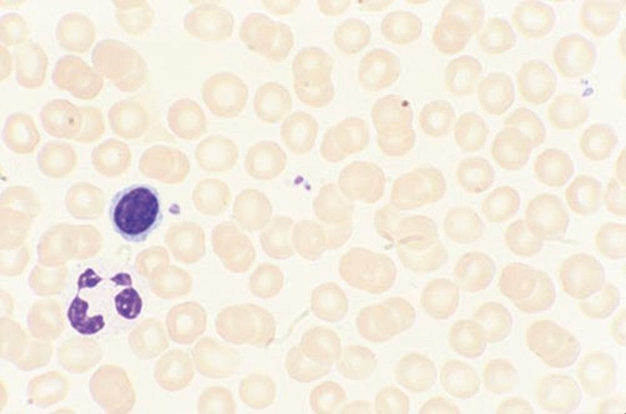
A drop of blood is applied against a glass slide that is subsequently stained with polychrome stains (Wright-Giemsa) to permit identification of the various cell types. These stains are mixtures of basic dyes (methylene blue) that stain as blue and acidic dyes (eosin) that stain as red. Thus, acid components of the cell (nucleus, cytoplasmic RNA, basophilic granules) stain blue or purple, and basic components of the cell (hemoglobin, eosinophilic granules) stain red or orange.
By permission of the publisher. From Tefferi A, Li C. In Atlas of Clinical Hematology. Edited by JO Armitage. Philadelphia, Current Medicine, 2004.
In these topics