


Various lesions can compress the spinal cord, causing segmental sensory, motor, reflex, and sphincter deficits. Diagnosis is by MRI. Treatment is directed at relieving compression.
(See also Overview of Spinal Cord Disorders and Immediate care for spinal trauma.)
Compression is caused far more commonly by lesions outside the spinal cord (extramedullary) than by lesions within it (intramedullary).
Compression may be
Acute
Subacute
Chronic
Acute compression develops within minutes to hours. It is often due to
Trauma (eg, vertebral crush fracture with displacement of fracture fragments, acute disk herniation, severe bone or ligamentous injury causing hematoma, vertebral subluxation or dislocation)
Metastatic tumor
It is occasionally due to abscess and rarely due to spontaneous epidural hematoma. Acute compression may follow subacute and chronic compression, especially if the cause is abscess or tumor.
Subacute compression develops over days to weeks. It is usually caused by
A metastatic extramedullary tumor
A subdural or an epidural abscess or hematoma
A cervical or, rarely, thoracic herniated disk
Chronic compression develops over months to years. It is commonly caused by
Bony protrusions into the cervical, thoracic, or lumbar spinal canal (eg, due to osteophytes or spondylosis, especially when the spinal canal is narrow, as occurs in spinal stenosis)
Compression can be aggravated by a herniated disk and hypertrophy of the ligamentum flavum. Less common causes include arteriovenous malformations and slow-growing extramedullary tumors.
Atlantoaxial subluxation and other craniocervical junction abnormalities may cause acute, subacute, or chronic spinal cord compression.
Lesions that compress the spinal cord may also compress nerve roots or, rarely, occlude the spinal cord’s blood supply, causing spinal cord infarction.
Symptoms and Signs of Spinal Cord Compression
Acute or advanced spinal cord compression causes segmental deficits, paraparesis or quadriparesis, hyporeflexia (when acute) followed by hyperreflexia, extensor plantar responses, loss of sphincter tone (with bowel and bladder dysfunction), and sensory deficits. Subacute or chronic compression may begin with local back pain, often radiating down the distribution of a nerve root (radicular pain), and sometimes hyperreflexia and loss of sensation. Sensory loss may begin in the sacral segments. Complete loss of function may follow suddenly and unpredictably, possibly resulting from secondary spinal cord infarction.
Spinal percussion tenderness is prominent if the cause is metastatic carcinoma, abscess, or hematoma.
Intramedullary lesions tend to cause poorly localized burning pain rather than radicular pain and to spare sensation in sacral dermatomes. These lesions usually result in spastic paresis.
Diagnosis of Spinal Cord Compression
MRI or CT myelography
Spinal cord compression is suggested by spinal or radicular pain with reflex, motor, or sensory deficits, particularly at a segmental level.
Pearls & Pitfalls
|

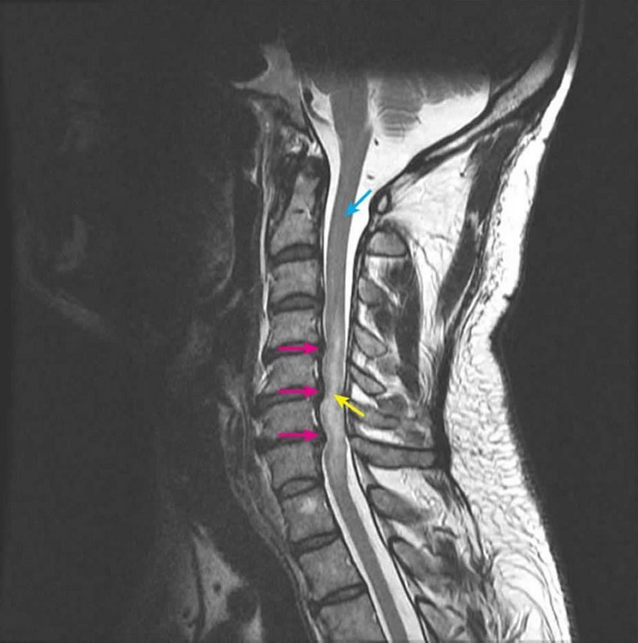
Courtesy of John Tsiouris, MD, Division of Neuroradiology, New York–Presbyterian Hospital/Weill Cornell Medical Center.
Treatment of Spinal Cord Compression
Relief of compression
Treatment of spinal cord compression is directed at relieving pressure on the cord. Incomplete or very recent complete loss of function may be reversible, but complete loss of function rarely is; thus, for acute compression, diagnosis and treatment must occur immediately.
Surgery is indicated in the following cases:
Neurologic deficits worsen despite nonsurgical treatment.
A biopsy is needed.
The spine is unstable.
Tumors recur after radiation therapy.
An abscess or a subdural or epidural hematoma is compressing the spinal cord.
Key Points
Spinal cord compression is usually secondary to an extrinsic mass.
Manifestations may include back and radicular pain (early) and segmental sensory and/or motor deficits, altered reflexes, extensor plantar responses, and loss of sphincter tone (with bowel and bladder dysfunction).
Do MRI or CT myelography immediately.
To relieve pressure on the cord, do surgery or give corticosteroids as soon as possible.


