


Cholestasis is failure of bilirubin secretion, resulting in conjugated hyperbilirubinemia and jaundice. There are numerous causes, which are identified by laboratory testing, hepatobiliary scan, and, sometimes, liver biopsy and surgery. Treatment depends on cause.
Cholestasis occurs in 1/2500 full-term infants. It is defined as direct bilirubin > 1 mg/dL (> 17.1 micromole/L).
Cholestasis is never normal and warrants evaluation.
Etiology of Neonatal Cholestasis
Cholestasis (see also Neonatal Hyperbilirubinemia) may result from extrahepatic or intrahepatic disorders, although some conditions overlap.
Extrahepatic causes of cholestasis
The most common extrahepatic disorder is
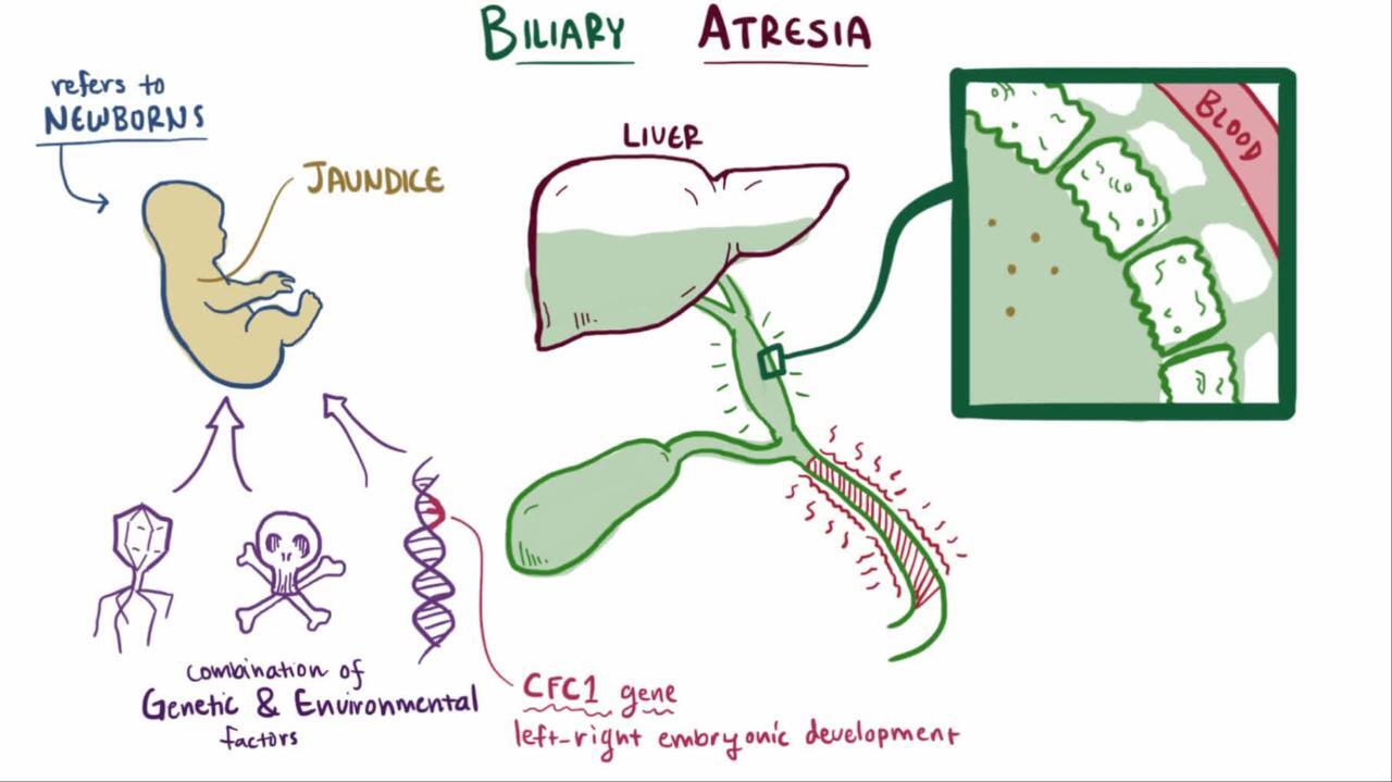
Biliary atresia (incidence in the United States about 1/8,000 to 1/18,000 live births) (1)
Biliary atresia is obstruction of the biliary tree due to progressive sclerosis of the extrahepatic bile duct. In most cases, biliary atresia manifests several weeks after birth, probably after inflammation and scarring of the extrahepatic (and sometimes intrahepatic) bile ducts. It is rarely present in preterm infants or in neonates at birth. The cause of the inflammatory response is unknown, but several infectious organisms have been implicated, including reovirus type 3 and cytomegalovirus.
Biliary cysts rarely manifest as neonatal cholestasis; these cysts are more common among patients with autosomal recessive polycystic kidney disease (2).
Inspissated bile duct syndrome can also be a cause of extrahepatic neonatal cholestasis and is more common among infants with cystic fibrosis.
Intrahepatic causes of cholestasis
Intrahepatic causes can be infectious, alloimmune, metabolic/genetic, or toxic.
Infections can cause cholestasis. Infections may be viral (eg, herpes simplex virus, cytomegalovirus, rubella), bacterial (eg, gram-positive and gram-negative bacteremia, urinary tract infection caused by Escherichia coli), or parasitic (eg, toxoplasmosis). Sepsis in neonates receiving parental nutrition can also cause cholestasis.
Gestational alloimmune liver disease involves transplacental passage of maternal IgG that induces a complement-mediated membrane attack complex that injures the fetal liver.
Metabolic causes include numerous inborn errors of metabolism such as galactosemia, tyrosinemia, alpha-1 antitrypsin deficiency, disorders of lipid metabolism, bile acid defects, mitochondrial disorders, and fatty acid oxidation defects. Additional genetic defects include Alagille syndrome, cystic fibrosis, and arthrogryposis-renal dysfunction-cholestasis (ARC) syndrome. There are also a number of gene mutations that interfere with normal bile production and excretion and cause cholestasis; the resultant disorders are termed progressive familial intrahepatic cholestasis.
Toxic causes are due mainly to the use of prolonged parenteral nutrition in extremely preterm neonates or infants with short bowel syndrome3).
Idiopathic neonatal hepatitis syndrome (giant cell hepatitis) is an inflammatory condition of the neonatal liver. Its incidence has decreased, and it is becoming rare because improved diagnostic studies allow identification of specific causes of cholestasis.
Etiology references
1. Harpavat S, Garcia-Prats JA, Anaya C, et al: Diagnostic yield of newborn screening for biliary atresia using direct or conjugated bilirubin measurements. JAMA 323(12):1141–1150, 2020. doi: 10.1001/jama.2020.0837
2. Fabris L, Fiorotto R, Spirli C, et al: Pathobiology of inherited biliary diseases: A roadmap to understand acquired liver diseases. Nat Rev Gastroenterol Hepatol 16(8):497–511, 2019. doi: 10.1038/s41575-019-0156-4
3. Guthrie G, Burrin D: Impact of Parenteral Lipid Emulsion Components on Cholestatic Liver Disease in Neonates. Nutrients 13(2):508, 2021. doi: 10.3390/nu13020508
Pathophysiology of Neonatal Cholestasis
In cholestasis, the primary failure is of bilirubin excretion, resulting in excess conjugated bilirubin in the bloodstream and decreased bile salts in the gastrointestinal (GI) tract. As a result of inadequate bile in the GI tract, there is malabsorption of fat and fat-soluble vitamins (A, D, E, and K), leading to vitamin deficiency, inadequate nutrition, and growth failure.
Symptoms and Signs of Neonatal Cholestasis
Cholestasis typically is noted in the first 2 weeks of life. Infants are jaundiced and often have dark urine (containing conjugated bilirubin), acholic stools, and hepatomegaly.
If cholestasis persists, chronic pruritus is common, as are symptoms and signs of fat-soluble vitamin deficiency; progression on growth charts may show a decline.
If the underlying disorder causes hepatic fibrosis and cirrhosis, portal hypertension with subsequent abdominal distention resulting from ascites, dilated abdominal veins, and upper GI bleeding resulting from esophageal varices may develop.
Diagnosis of Neonatal Cholestasis
Total and direct bilirubin
Liver tests
Tests for metabolic, infectious, and genetic causes
Liver ultrasonography
Hepatobiliary scan
Occasionally biopsy of liver, operative cholangiography, or genetic testing
Any infant who is jaundiced after age 2 weeks should be evaluated for cholestasis including with total and direct bilirubin levels. Some experts advocate that breastfed infants who have jaundice do not need to be evaluated until age 3 weeks. The initial approach should be directed at diagnosing treatable conditions (eg, extrahepatic biliary atresia, in which early surgical intervention improves short-term outcome).
Once cholestasis is confirmed, testing is required to determine etiology (see table Diagnostic Evaluation for Neonatal Cholestasis) and evidence of malabsorption (eg, low levels of the fat-soluble vitamins E, D, K, and A, or prolonged PT, suggesting a low level of vitamin K).
Abdominal ultrasonography is often the first test; it is noninvasive and can assess liver size and certain abnormalities of the gallbladder and common bile duct. However, it is nonspecific. A hepatobiliary scan using hydroxy iminodiacetic acid (HIDA scanERCP) can be done to investigate the anatomy of the biliary ductal system.
When no diagnosis has been made, a liver biopsy is generally done relatively early on, sometimes with operative cholangiography. Patients with biliary atresia typically have enlarged portal triads, bile duct proliferation, and increased fibrosis. Neonatal hepatitis is characterized by lobular disarray with multinucleated giant cells. Alloimmune liver disease is characterized by elevated hepatic iron stores.
Treatment of Neonatal Cholestasis
Specific cause treated
Medium-chain triglycerides
Sometimes ursodeoxycholic acid
For suspected biliary atresia, surgical exploration and sometimes portoenterostomy
Specific treatment is directed at the cause. If there is no specific therapy, treatment is supportive and consists primarily of nutritional therapy, including supplements of vitamins A, D, E, and K.
For formula-fed infants, a formula that is high in medium-chain triglycerides should be used because it is absorbed better in the presence of bile salt deficiency. Adequate calories are required; infants may need >
Cholestasis caused by parenteral nutrition resolves if the parenteral nutrition is stopped or sometimes if a newer-generation lipid emulsion is substituted for an older one before the infant develops severe liver disease.
Because gestational alloimmune liver disease has no definitive marker and/or test, treatment with IV immune globulin (IVIG) or exchange transfusion needs to be considered early to reverse the ongoing liver injury if no definite diagnosis has been made (1).
Treatment reference
1. Fischer HS, Staufner C, Sallmon H, et al: Early exchange transfusion to treat neonates with gestational alloimmune liver disease: An 11-year cohort study. J Pediatr Gastroenterol Nutr 70(4):444–449, 2020. doi: 10.1097/MPG.0000000000002593
Prognosis for Neonatal Cholestasis
Biliary atresia is progressive and, if untreated, results in liver failure, cirrhosis with portal hypertension by several months of age, and death by 1 year of age.
Prognosis for cholestasis due to specific disorders (eg, metabolic disease) is variable, ranging from a completely benign course to a progressive disease resulting in cirrhosis.
Idiopathic neonatal hepatitis syndrome usually resolves slowly, but permanent liver damage may result and lead to liver failure and death.
Gestational alloimmune liver disease has a poor prognosis without early intervention.
Key Points
There are numerous inherited and acquired causes of neonatal cholestasis, resulting in failure of bilirubin excretion and thus excess conjugated bilirubin.
Neonatal cholestasis typically is noted in the first 2 weeks of life; infants are jaundiced and often have dark urine, acholic stools, and hepatomegaly.
Begin with laboratory testing of the liver, ultrasonography, and hepatobiliary scan and do tests for causes, sometimes including liver biopsy.
Treat specific cause and give supportive care, including supplementation of fat-soluble vitamins and a formula that is high in medium-chain triglycerides and contains sufficient calories.
More Information
The following English-language resources may be useful. Please note that THE MANUAL is not responsible for the content of these resources.
North American Society for Pediatric Gastroenterology, Hepatology, and Nutrition and the European Society for Pediatric Gastroenterology, Hepatology, and Nutrition: Guideline for the evaluation of cholestatic jaundice in infants (2017)
North American Society for Pediatric Gastroenterology, Hepatology, and Nutrition and the European Society for Pediatric Gastroenterology, Hepatology, and Nutrition: Joint position paper on nutritional support of children with chronic liver diseases (2019)


