


Hydrocephalus is an accumulation of extra fluid in the normal spaces within the brain (ventricles) and/or between the inner and middle layers of tissues that cover the brain (the subarachnoid space). The extra fluid usually causes an enlarged head and developmental problems.
Hydrocephalus occurs when the fluid in the normal spaces in the brain (ventricles) cannot drain.
The fluid may accumulate for many reasons, such as a birth defect, bleeding within the brain, or a brain tumor.
Typical symptoms include an abnormally large head and abnormal development.
The diagnosis is based on computed tomography (CT), ultrasonography, or magnetic resonance imaging (MRI).
Surgery is needed to insert a drain (shunt) into the brain or to create an opening that allows fluid to drain.
(See also Overview of Brain and Spinal Cord Birth Defects.)
The fluid surrounding the brain is called cerebrospinal fluid. This fluid is produced in spaces within the brain called ventricles. The fluid is constantly being produced and must drain to a different area, where it is absorbed into the blood. When the fluid cannot drain, it accumulates in the ventricles and/or subarachnoid space, causing hydrocephalus (water on the brain). Often, the pressure in the ventricles and within the brain increases, compressing brain tissue.
Many conditions, such as a birth defect, bleeding within the brain (which is a complication particularly for premature infants), or brain tumors, can block drainage and cause hydrocephalus. Hydrocephalus may also be caused by certain gene defects.
Infants can be born with hydrocephalus or it can occur during or after birth.

STEVE ALLEN/SCIENCE PHOTO LIBRARY
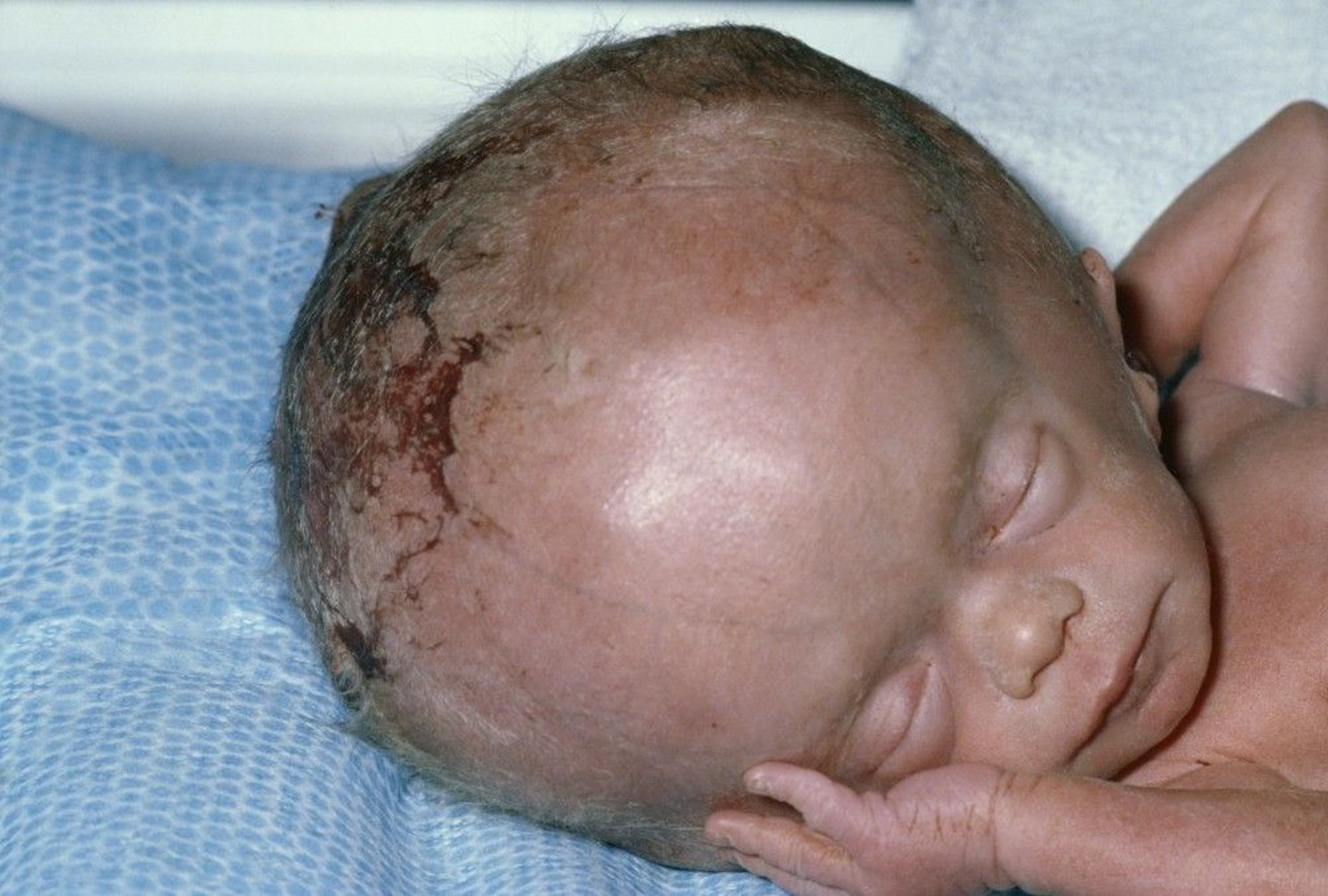
Symptoms of Hydrocephalus
An abnormally large head may be a sign of hydrocephalus.
When pressure in the brain is increased because of hydrocephalus, infants are irritable and listless, have a high-pitched cry, and vomit, and they may have seizures. Also, the soft spots between the skull bones (called the fontanelles) may bulge, causing a soft bump on the head. The eyes may not move together, sometimes making the child look cross-eyed (called strabismus).
Older children may have a headache, problems with vision, or both.
If hydrocephalus is not treated, infants do not develop normally. Some children with hydrocephalus, especially those who develop hydrocephalus early in the pregnancy, are intellectually disabled or have learning disabilities, a seizure disorder (epilepsy), or, in girls, early puberty. Some children have vision loss.
Other children develop normal intelligence.
Diagnosis of Hydrocephalus
Before birth, prenatal ultrasonography
After birth, computed tomography, magnetic resonance imaging, or ultrasonography of the head
Before birth, hydrocephalus is often detected when routine prenatal ultrasonography is done.
After birth, doctors suspect the diagnosis in newborns based on symptoms noted during a routine physical examination. Doctors then do ultrasonography of the head to confirm the diagnosis of hydrocephalus.
In older infants and children, doctors do computed tomography (CT), magnetic resonance imaging (MRI), or ultrasonography of the head to confirm the diagnosis.
Once the diagnosis has been made, all children undergo CT or ultrasonography to monitor the hydrocephalus and determine whether it is worsening.
Treatment of Hydrocephalus
Sometimes spinal taps
For worsening hydrocephalus, a shunt or opening in the ventricles
The goal of treatment is
To keep pressure within the brain normal

Treatment of hydrocephalus depends on what is causing the disorder, how severe it is, and whether it is worsening.
If needed, pressure within the brain can often be temporarily reduced by removing spinal fluid with repeated spinal taps (lumbar punctures) until a shunt is placed.
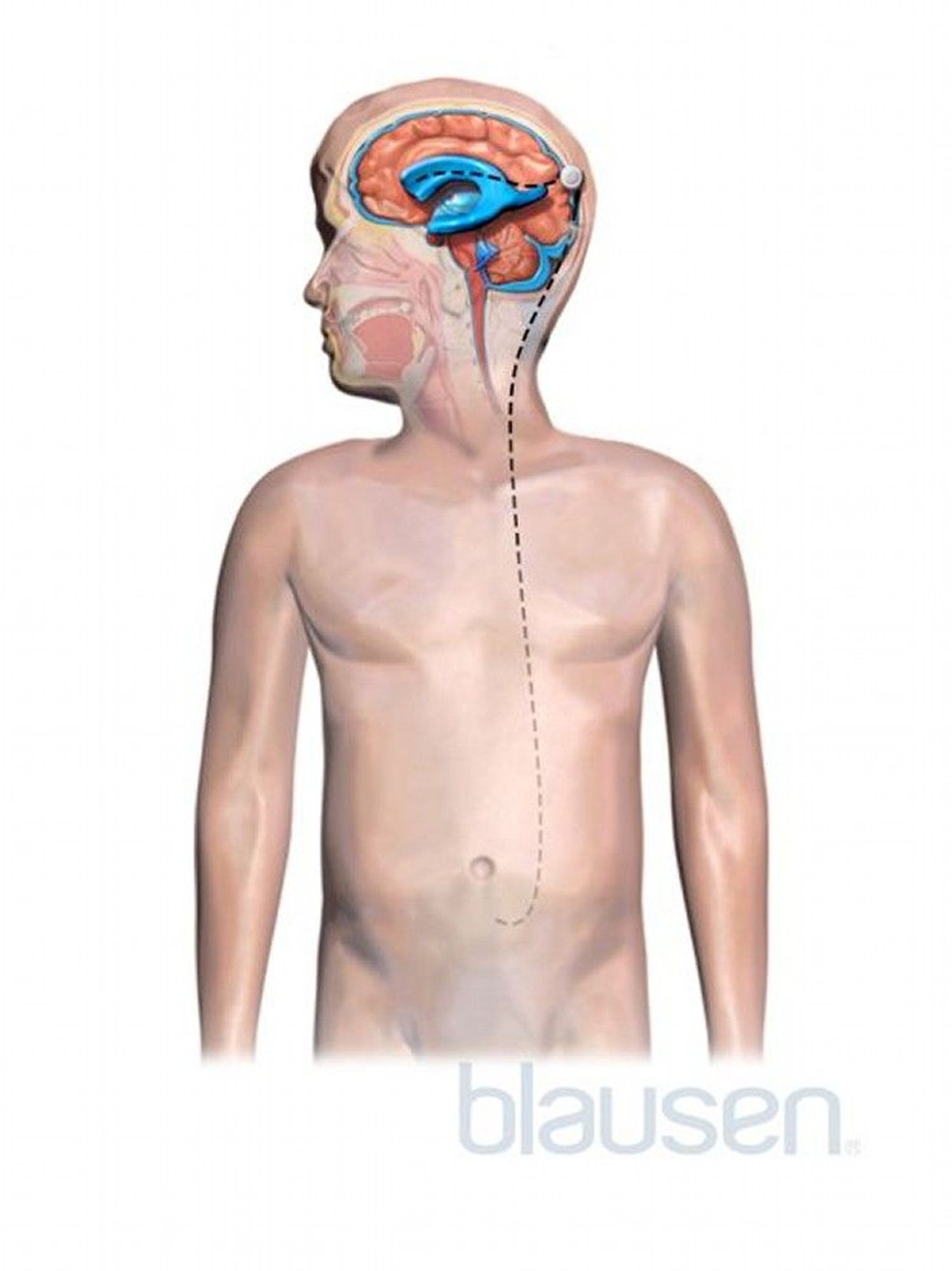
If hydrocephalus is worsening, doctors place a ventricular shunt. The shunt is a plastic tube that creates a permanent alternate drainage path for cerebrospinal fluid. Draining the cerebrospinal fluid decreases the pressure and volume of the fluid inside the brain. Doctors place the shunt in the ventricles in the brain and run it under the skin from the head to another site, usually the abdomen (called a ventriculoperitoneal shunt, or VP shunt). The shunt contains a valve that allows fluid to leave the brain if the pressure becomes too high.
Although a few children can eventually do without the shunt as they get older, shunts are rarely removed because of the risk of bleeding and injury.
In some children, doctors do a ventriculostomy. In this procedure, doctors do not place a shunt and instead create an opening between a ventricle and the subarachnoid space in the brain. This opening allows excess fluid to drain and be absorbed as usual. Sometimes a shunt is still necessary if the ventriculostomy does not cure the hydrocephalus.
After placing a shunt or doing a ventriculostomy, doctors measure head circumference and determine how the child is developing. Imaging tests (such as computed tomography or magnetic resonance imaging) are done periodically.
Complications of shunts
Shunts can become infected. Children who develop an infection are given antibiotics. Typically the shunt is then removed and replaced.
Shunts can break or become blocked and stop functioning correctly. To determine how a shunt is functioning, doctors take x-rays of the shunt and do imaging tests of the brain. A shunt that is not working correctly is typically removed and replaced.
More Information
The following English-language resource may be useful. Please note that THE MANUAL is not responsible for the content of this resource.
March of Dimes: An organization for pregnant people and babies that provides support and information about how to prevent maternal health risks, premature birth, and mother and infant deaths


