


Impetigo is a superficial skin infection with crusting or bullae caused by streptococci, staphylococci, or both. Ecthyma is an ulcerative form of impetigo. Diagnosis is clinical. Treatment is with topical and sometimes oral antibiotics.
(See also Overview of Bacterial Skin Infections.)
No predisposing lesion is identified in most patients, but impetigo may follow any type of break in the skin. General risk factors seem to be a moist environment, poor hygiene, or chronic nasopharyngeal carriage of staphylococci or streptococci.
Impetigo may be bullous or nonbullous. Staphylococcus aureus is the predominant cause of nonbullous impetigo and the cause of all bullous impetigo. Bullae are caused by exfoliative toxin produced by staphylococci. Methicillin-resistant S. aureus (MRSA) has been isolated in many cases of impetigo.
Symptoms and Signs of Impetigo and Ecthyma
Nonbullous impetigo typically manifests as clusters of vesicles or pustules that rupture and develop a honey-colored crust (exudate from the lesion base) over the lesions. Smaller lesions may coalesce into larger crusted plaques.
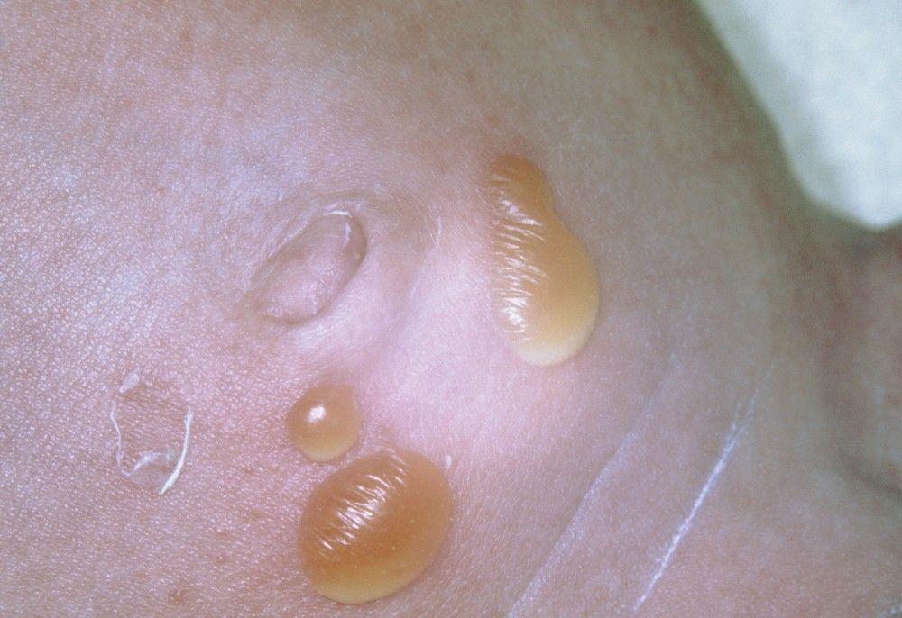
Bullous impetigo is similar except that vesicles typically enlarge rapidly to form bullae. The bullae burst and expose larger bases, which become covered with honey-colored varnish or crust.
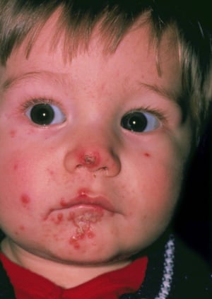
DR P. MARAZZI/SCIENCE PHOTO LIBRARY
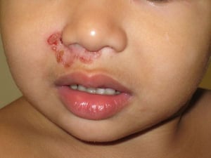
Image courtesy of Wingfield Rehmus, MD, MPH.
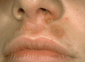
Image provided by Thomas Habif, MD.
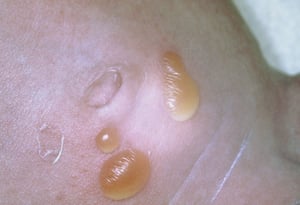
SCIENCE PHOTO LIBRARY

Image provided by Thomas Habif, MD.


DR P. MARAZZI/SCIENCE PHOTO LIBRARY

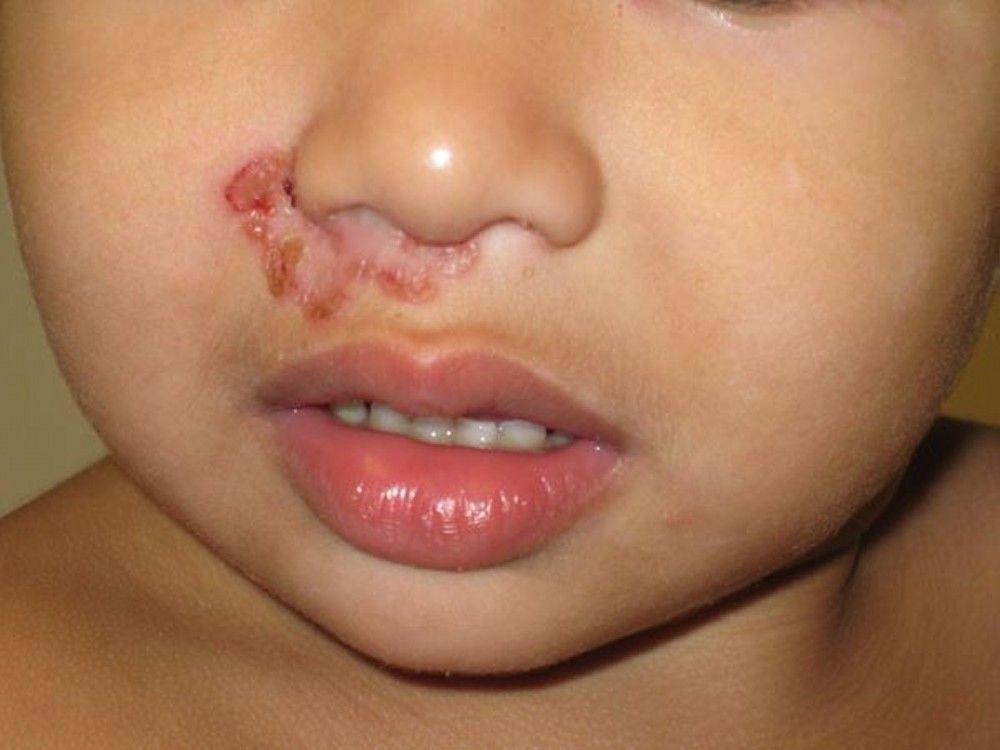
Image courtesy of Wingfield Rehmus, MD, MPH.

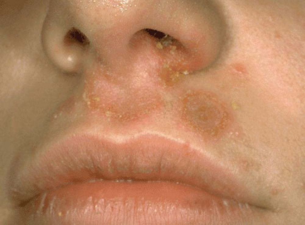
Image provided by Thomas Habif, MD.

SCIENCE PHOTO LIBRARY

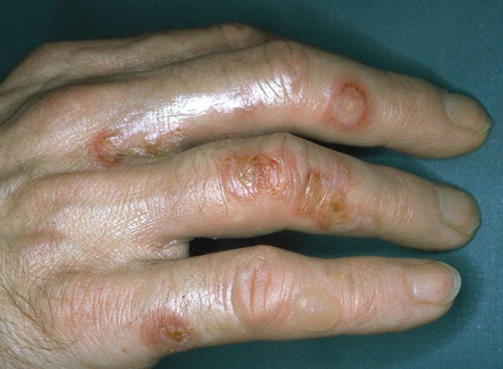
Image provided by Thomas Habif, MD.
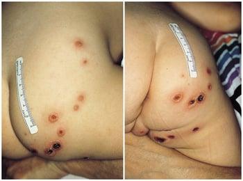
Ecthyma is a form of impetigo characterized by small, purulent, shallow, punched-out ulcers with thick, brown-black crusts and surrounding erythema.

Impetigo and ecthyma cause mild pain or discomfort. Pruritus is common; scratching may spread infection, inoculating adjacent and nonadjacent skin.
Diagnosis of Impetigo and Ecthyma
Clinical evaluation
Diagnosis of impetigo and ecthyma is by characteristic appearance.
Cultures of lesions are indicated only when the patient does not respond to empiric therapy. Patients with recurrent impetigo should have nasal culture. Persistent infections should be cultured to identify MRSA.
Treatment of Impetigo and Ecthyma
Topical mupirocin, retapamulin, fusidic acid, or ozenoxacin
Sometimes oral antibiotics
The affected area should be washed gently with soap and water several times a day to remove any crusts.
Use of initial empiric therapy against MRSA
Other therapy includes restoring a normal cutaneous barrier in patients with underlying atopic dermatitis or extensive xerosis
Prompt recovery usually follows timely treatment. Delay can cause cellulitis, lymphangitis, furunculosis, and hyperpigmentation or hypopigmentation with or without scarring. Children aged 2 to 4 years are at risk of acute glomerulonephritis if nephritogenic strains of group A streptococci are involved (types 49, 55, 57, and 59); nephritis seems to be more common in the southern United States than in other regions. It is unlikely that treatment with antibiotics prevents poststreptococcal glomerulonephritis.
Key Points
Staphylococcus aureus causes most nonbullous impetigo and all bullous impetigo.
Honey-colored crust is characteristic of bullous and nonbullous impetigo.
For persistent impetigo, culture the lesion (to identify methicillin-resistant S. aureus [MRSA]) and the nose (to identify a potential nasal reservoir).
Treat most cases with topical antibiotics.


