


Gallbladder and bile duct tumors can cause extrahepatic biliary obstruction. Symptoms may be absent but often are constitutional or reflect biliary obstruction. Diagnosis is based on ultrasonography plus CT cholangiography or magnetic resonance cholangiopancreatography. Prognosis is grim. Mechanical bile drainage can often relieve pruritus, recurrent sepsis, and pain due to biliary obstruction.
(See also Overview of Biliary Function.)
Cholangiocarcinomas and other bile duct tumors are rare (1 to 2/100,000 people) but are usually malignant (1). Cholangiocarcinomas occur predominantly in the extrahepatic bile ducts: 60 to 70% in the perihilar region (Klatskin tumors), about 25% in the distal extrahepatic ducts, and the rest in the liver. Established risk factors include primary sclerosing cholangitis, older age, infestation with liver flukes, and a choledochal cyst.
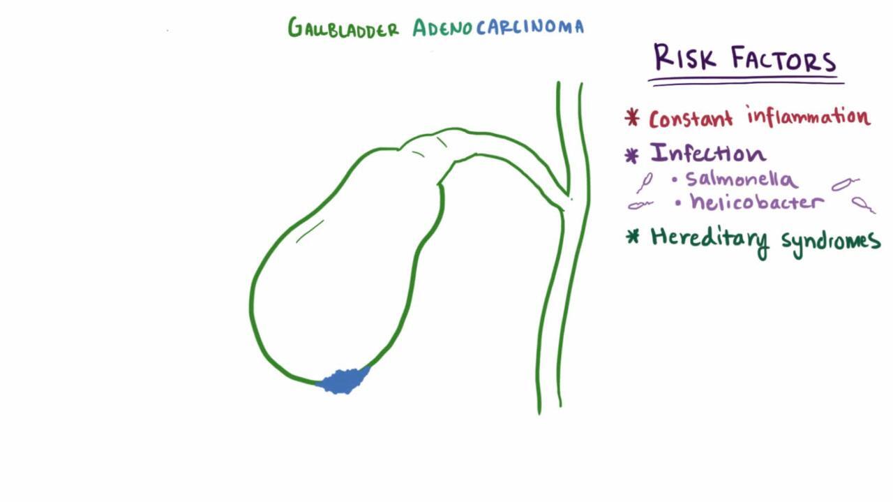
Gallbladder carcinoma is uncommon (2.5/100,000). It is more common among American Indians, patients with large gallstones (> 3 cm), and those with extensive gallbladder calcification due to chronic cholecystitis (porcelain gallbladder). Nearly all (70 to 90%) patients also have gallstones. Median survival is 3 months. Cure is possible when cancer is found early (eg, incidentally at cholecystectomy).
Gallbladder polyps are usually asymptomatic benign mucosal projections that develop in the lumen of the gallbladder. Most are < 10 mm in diameter and composed of cholesterol ester and triglycerides; the presence of such polyps is called cholesterolosis. They are found in about 5% of people during ultrasonography. Other, much less common benign polyps include adenomas (causing adenomyomatosis) and inflammatory polyps. Patients with gallbladder polyps < 10 mm should undergo surveillance ultrasound every 6 to 12 months based on their risk factors and polyp size. Cholecystectomy is sometimes recommended with some polyps 6 to 9 mm in diameter. However, if diameter is >10 mm, surgery should always be considered.
General reference
1. Banales JM, Cardinale V, Carpino G, et al: Expert consensus document: Cholangiocarcinoma: current knowledge and future perspectives consensus statement from the European Network for the Study of Cholangiocarcinoma (ENS-CCA). Nat Rev Gastroenterol Hepatol 13(5):261-280, 2016. doi: 10.1038/nrgastro.2016.51
Symptoms and Signs of Gallbladder and Bile Duct Tumors
Most patients with cholangiocarcinomas present with pruritus and painless obstructive jaundice, typically at age 50 to 70 years. Early perihilar tumors may cause only vague abdominal pain, anorexia, and weight loss. Other features include fatigue, acholic stool, a palpable mass, hepatomegaly, or a distended gallbladder (Courvoisier sign, with distal cholangiocarcinoma). Pain may resemble that of biliary colic (reflecting biliary obstruction) or may be constant and progressive. Sepsis (secondary to acute cholangitis), although unusual, may be induced by endoscopic retrograde cholangiopancreatography (ERCP).
Manifestations of gallbladder carcinoma may range from incidental findings at cholecystectomy done to relieve biliary pain to cholelithiasis to advanced disease with constant pain, weight loss, and an abdominal mass or obstructive jaundice.
Most gallbladder polyps cause no symptoms.
Diagnosis of Gallbladder and Bile Duct Tumors
Ultrasonography (sometimes endoscopic), followed by magnetic resonance cholangiopancreatography (MRCP) or CT cholangiography
Sometimes endoscopic retrograde cholangiopancreatography (ERCP)
Cholangiocarcinomas and gallbladder carcinomas are suspected when extrahepatic biliary obstruction is unexplained. Laboratory test results reflect the degree of cholestasis. In patients with primary sclerosing cholangitis, serum carcinoembryonic antigen (CEA) and carbohydrate antigen (CA) levels 19-9 are measured periodically to check for cholangiocarcinoma.
Diagnosis is based on ultrasonography (or endoscopic ultrasonography), typically followed by MRCP (see Imaging Tests of the Liver and Gallbladder). CT is sometimes done and may provide more information than ultrasonography, particularly for gallbladder carcinomas. When these methods are inconclusive or if cholangiocarcinoma is suspected, ERCP is necessary. ERCP not only detects the tumor but also, with brushings, can provide a tissue diagnosis, sometimes making ultrasonography- or CT-guided needle biopsy unnecessary. Contrast-enhanced CT assists in staging.
Open laparotomy is necessary to determine disease extent, which guides treatment.
Treatment of Gallbladder and Bile Duct Tumors
For cholangiocarcinomas, stenting (or another bypass procedure) or occasionally resection
For gallbladder carcinoma, usually symptomatic treatment
For cholangiocarcinoma, stenting or surgically bypassing the obstruction relieves pruritus, jaundice, and perhaps fatigue.
Hilar cholangiocarcinomas with CT evidence of spread are stented via percutaneous transhepatic cholangiography or endoscopic retrograde cholangiopancreatography (ERCP). Distal duct cholangiocarcinomas are stented endoscopically with ERCP. If cholangiocarcinoma appears localized, surgical exploration determines resectability by hilar resection or pancreaticoduodenectomy.
Liver transplantation for localized hilar cholangiocarcinoma is available at some transplant centers as part of a specific protocol approved by the United Network for Organ Sharing (UNOS).
Many gallbladder carcinomas are treated symptomatically.
Key Points
Biliary tract cancer (usually cholangiocarcinoma or gallbladder carcinoma) is uncommon.
Suspect cancer if patients have an unexplained extrahepatic biliary obstruction or abdominal mass.
Diagnose cancers by imaging, beginning with ultrasonography, followed by MRCP.
Treat cancers symptomatically (eg, by stenting or bypassing obstructions in cholangiocarcinoma); occasionally, resection is warranted.
Consider liver transplantation for selected patients with hilar cholangiocarcinoma.


