


Immunotactoid Glomerulopathy

Immunotactoid Glomerulopathy
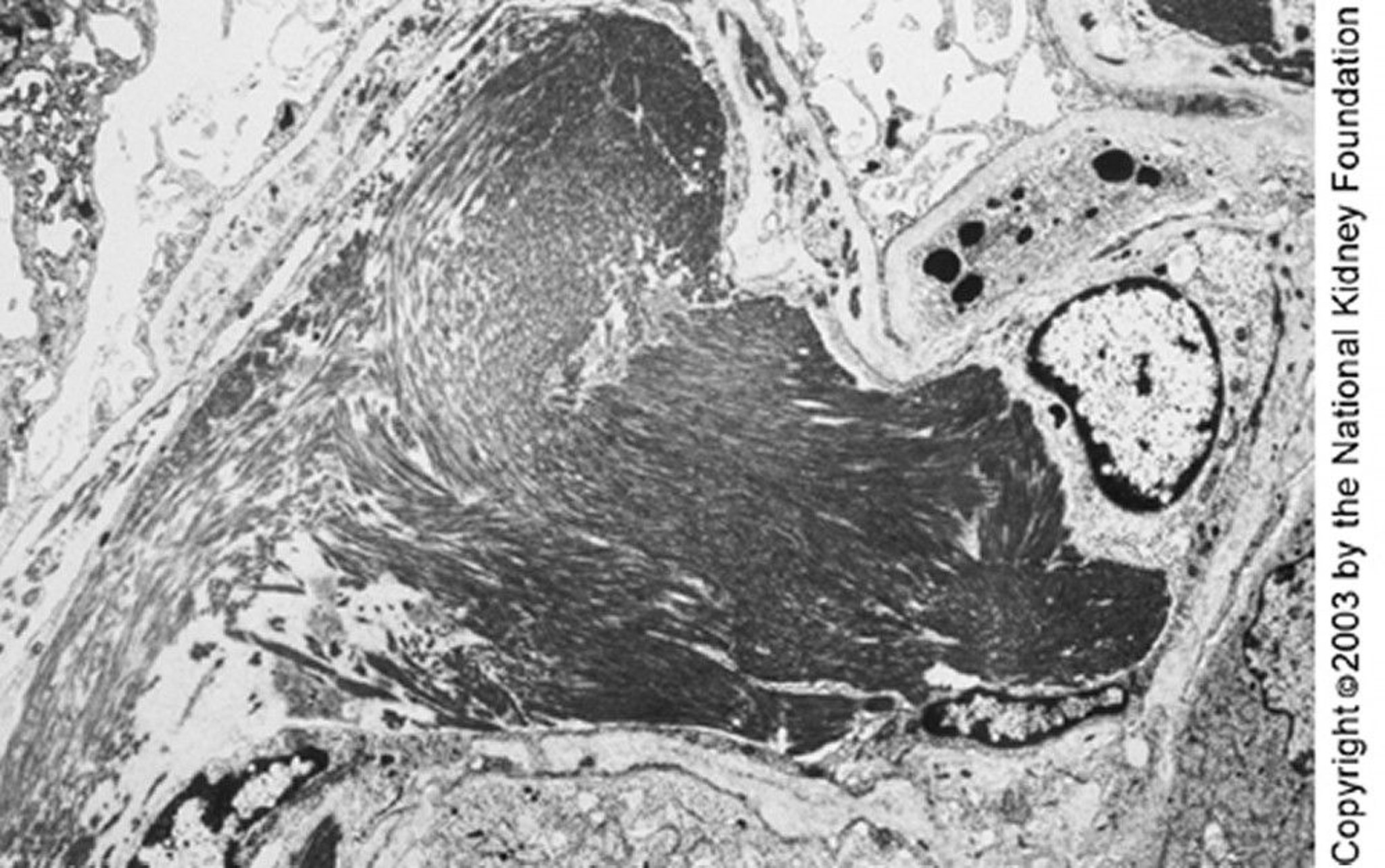
Large microtubules (close to 50 nm in diameter) organized in parallel arrays can be seen on transmission electron microscopy. The parallel deposits and microtubular structure may distinguish immunotactoid from fibrillary glomerulonephritis (×4000).
Image provided by Agnes Fogo, MD, and the American Journal of Kidney Diseases' Atlas of Renal Pathology (see www.ajkd.org).
In these topics