


Compressive Myelopathy

Compressive Myelopathy
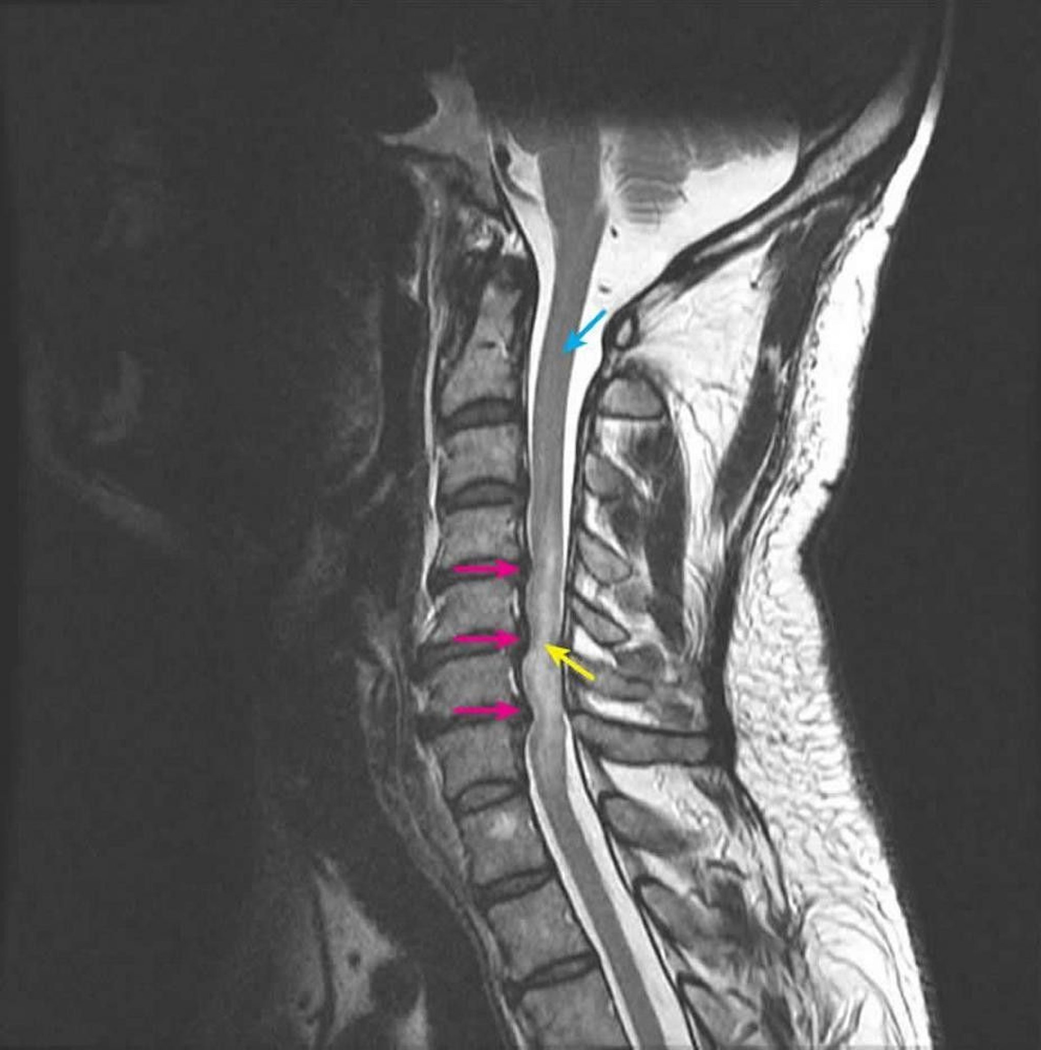
Sagittal T2-weighted MRI shows spinal cord compression at the C4-5, C5-6, and C6-7 intervertebral levels secondary to multilevel posterior disk herniations (pink arrows). The intramedullary T2 signal is abnormally increased, representing spinal cord edema (yellow arrow). The normal spinal cord signal is seen at the C2 level (blue arrow).
Courtesy of John Tsiouris, MD, Division of Neuroradiology, New York–Presbyterian Hospital/Weill Cornell Medical Center.
In these topics