


Craniotubular hyperostoses are osteopetroses that involve bony overgrowths that alter contour and increase skeletal density.
Diaphyseal dysplasia (Camurati-Engelmann disease)
This autosomal dominant disorder is caused by mutations in the TGFB1 gene.
Diaphyseal dysplasia manifests during mid-childhood with muscular pain, weakness, and wasting, typically in the legs. These symptoms usually resolve by age 30 years. Hyperostoses affect the long bones and skull. Cranial nerve compression and elevated intracranial pressure occur occasionally. Some patients are severely handicapped; others are virtually asymptomatic.
Diagnosis of diaphyseal dysplasia is suspected by the combination of muscular deficits and hyperostoses of the long bones and skull. Typically, plain x-rays are done. The predominant x-ray feature is marked thickening of the periosteal and medullary surfaces of the diaphyseal cortices of the long bones, but findings vary. Medullary canals and external bone contours are irregular. The extremities and axial skeleton usually are spared. Rarely, the skull is involved, with calvarial widening and basal sclerosis.
Corticosteroids may help relieve bone pain and improve muscle strength.
Endosteal hyperostosis (van Buchem syndrome)
This disorder is usually autosomal recessive. In endosteal hyperostosis, genetic lesions seem to affect the normal function of osteoblasts.
Overgrowth and distortion of the mandible and brow become evident during mid-childhood. Subsequently, cranial nerves become entrapped, leading to facial palsy and deafness. Life span is not compromised, stature is normal, and bones are not fragile.
X-rays show widening and sclerosis of the calvaria, cranial base, and mandible. Diaphyseal endosteum in the tubular bones is thickened.
Surgical decompression of entrapped nerves may be helpful.
Sclerosteosis
This autosomal recessive disorder is caused by a mutation in the SOST gene, which codes for the protein sclerostin. Sclerosteosis is most common among Afrikaners of South Africa.
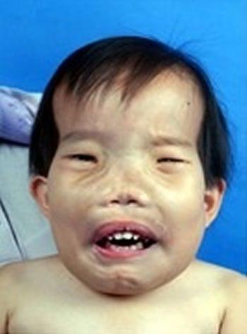
Overgrowth and sclerosis of the skeleton, particularly of the skull, develop during early childhood. Height and weight are often excessive. Initial symptoms and signs of sclerosteosis may include deafness and facial palsy due to cranial nerve entrapment. Distortion of facies, apparent by age 10 years, eventually becomes severe. Cutaneous or bony syndactyly of the second and third fingers distinguishes sclerosteosis from other forms of craniotubular hyperostoses.

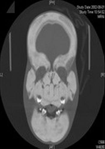
Diagnosis of sclerosteosis is suspected by characteristic skeletal abnormalities, particularly when the patient also has syndactyly. Typically, plain x-rays are done. Predominant x-ray features are gross widening and sclerosis of the calvaria and mandible. Vertebral bodies are spared, although their pedicles are dense. Pelvic bones are sclerotic but have normal contours. Long bones have sclerosed, hyperostotic cortices and undermodeled shafts. A diagnostic genetic test is available.

Surgery to relieve intracranial pressure or to decompress entrapped nerves may help.


