


Retinoblastoma is a cancer of the retina that occurs almost exclusively in children < 2 years old. Symptoms and signs commonly include leukocoria (a white reflex in the pupil), strabismus, and, less often, inflammation and impaired vision. Diagnosis is based on ophthalmoscopic examination and ultrasonography, CT, or MRI. Treatment of small cancers and bilateral disease may include photocoagulation, cryotherapy, and radiation therapy. Treatment of advanced and some larger cancers is enucleation. Chemotherapy is sometimes used to reduce cancer volume and to treat cancers that have spread beyond the eye.
Retinoblastoma occurs in 1/15,000 to 1/30,000 live births and represents about 2% of childhood cancers (1). It is usually diagnosed in children < 2 years of age; < 5% of cases are diagnosed in those > 5 years of age.
The cancer may be hereditary; inheritance is mainly autosomal dominant but with incomplete penetrance (clinical symptoms are not always present in individuals who have the disease-causing mutation). About 25% of patients have bilateral disease, which is always heritable. Another 15% of patients have heritable unilateral disease, and the remaining 60% have nonhereditary unilateral disease.
The pathogenesis of inheritance appears to involve mutational deactivation of both alleles of a retinoblastoma suppressor gene (RB1) located on chromosome 13q14. In the hereditary form, a germline mutation alters one allele in all cells, and a later somatic mutation alters the other allele in the retinal cells (the second hit in this two-hit model), resulting in the cancer. The nonhereditary form probably involves somatic mutation of both alleles in a retinal cell. Although the term "sporadic" can be used to describe nonhereditary forms of retinoblastoma, it is technically a misnomer because many sporadic cases are due to de novo germline mutations and are therefore also subsequently heritable.
General reference
1. Siegel RL, Miller KD, Fuchs HE, et al: Cancer Statistics, 2022. CA Cancer J Clin 72(1):7–33, 2022. doi: 10.3322/caac.21708
Symptoms and Signs of Retinoblastoma
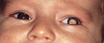
Patients typically present with leukocoria (a white reflex in the pupil, sometimes referred to as cat’s-eye pupil) or strabismus.
Much less often, patients present with inflammation of the eye or impaired vision.

Rarely, the cancer has already spread, via the optic nerve or the choroid or hematogenously, resulting in an orbital or soft-tissue mass, local bone pain, headache, anorexia, or vomiting.
Diagnosis of Retinoblastoma
Orbital ultrasonography, CT, or MRI
Sometimes optical coherence tomography, bone scan, bone marrow aspirate and biopsy, and lumbar puncture
When the diagnosis is suspected, both fundi must be closely examined by indirect ophthalmoscopy with the pupils widely dilated and the child under general anesthesia. The cancers appear as single or multiple gray-white elevations in the retina; cancer seeds may be visible in the vitreous.
Diagnosis of retinoblastoma is usually confirmed by orbital ultrasonography, MRI, or CT. In almost all cancers, calcification can be detected by CT. However, if the optic nerve appears abnormal during ophthalmoscopy, orbital MRI is better for finding cancer extension into the optic nerve or choroid. Optical coherence tomography, a noninvasive imaging test, is sometimes used.
If optic nerve extension is suspected or extensive choroidal invasion is present, a lumbar puncture and brain MRI should be done to assess for metastasis. Because distant metastasis is rare, bone marrow evaluation and bone scan can be reserved for patients with bony symptoms or for those with clear evidence of metastatic disease.
Because of the risk of tumor spread, there is no clear role for ocular tumor biopsy to obtain a tissue diagnosis.
Patients with retinoblastoma require molecular genetic testing, and if a germline mutation is identified, parents should also be tested for the same mutation. If subsequent offspring of parents have the germline mutation, the same genetic testing and regular ophthalmologic examination are required. Recombinant DNA probes may be useful for detecting asymptomatic carriers.
Children who have a parent or sibling with a history of retinoblastoma should be evaluated by an ophthalmologist shortly after birth and then every 4 months until age 4 years.
Treatment of Retinoblastoma
For advanced unilateral cancer, enucleation
For less advanced unilateral cancer, sometimes chemotherapy and/or local-control treatments
For bilateral cancer, photocoagulation, intra-arterial chemotherapy, or unilateral enucleation with photocoagulation, cryotherapy and irradiation of the other eye
Systemic chemotherapy
The goal of retinoblastoma treatment should be cure, but attempts to preserve as much vision as possible are appropriate. The treatment approach depends on the size of the tumor (more or less than 3 mm in dimension/thickness), spread to surrounding areas, and functionality of the eye. A multidisciplinary team is highly recommended and should include a pediatric ophthalmologist with expertise in retinoblastoma, a pediatric oncologist, and a radiation oncologist.
Advanced unilateral retinoblastoma (large tumor with evidence of disease extension) is managed by enucleation with removal of as much of the optic nerve as possible. In less advanced cases where preservation of vision is a possibility, conservative eye salvage approaches with chemotherapy and/or local-control treatments can be considered.
For patients with bilateral cancer, vision can usually be preserved. Options include bilateral photocoagulation, intra-arterial chemotherapy, or unilateral enucleation and photocoagulation, cryotherapy, and irradiation of the other eye. Radiation therapy is by external beam or, for very small cancers, brachytherapy (attachment of a radioactive plaque to the eye wall near the cancer).
Ophthalmologic re-examination of both eyes and retreatment, if necessary, are required at 2- to 4-month intervals.
Prognosis for Retinoblastoma
If the cancer is treated when it is intraocular, > 90% of patients can be cured. Prognosis for patients with metastatic disease is poor (1).
In patients with hereditary retinoblastoma, incidence of second cancers is increased; about 50% arise within the irradiated area. These cancers can include sarcomas and malignant melanoma. About 70% of patients who will develop a second cancer develop it within 30 years of the primary retinoblastoma.
Prognosis reference
1. Dunkel IJ, Piao J, Chantada GL, et al: Intensive multimodality therapy for extraocular retinoblastoma: A Children's Oncology Group trial (ARET0321). J Clin Oncol 40(33):3839–3847, 2022. doi: 10.1200/JCO.21.02337


