


Bronchoscopy is the introduction of an endoscope into the airways.
Flexible fiberoptic bronchoscopy (rather than rigid bronchoscopy) is used for virtually all diagnostic, and most therapeutic, indications.

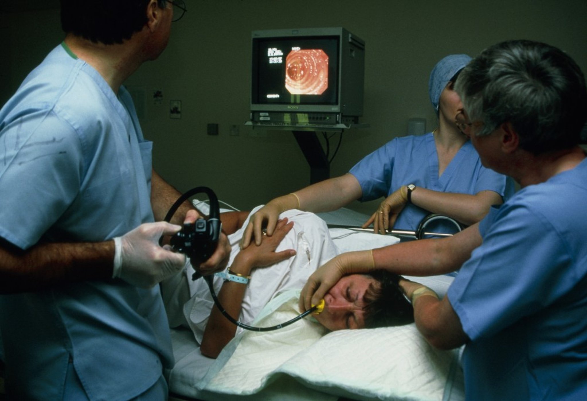
ANTONIA REEVE/SCIENCE PHOTO LIBRARY
Indications for Bronchoscopy
Flexible bronchoscopes facilitate airway visualization and documentation of findings (see table Indications for Flexible Fiberoptic Bronchoscopy).
Diagnostically, flexible fiberoptic bronchoscopy allows for
Direct airway visualization down to, and including, subsegmental bronchi
Sampling of respiratory secretions and cells via bronchial washings, brushings, and lavage of peripheral airways and alveoli
Biopsy of endobronchial, parenchymal, and mediastinal structures
Therapeutic uses include
Suctioning of retained secretions
Placing an endobronchial tube, stent, or valve
Removing foreign objects
Using balloon dilation to relieve airway stenoses
Rigid bronchoscopy is used only when a wider aperture and channels are required for better visualization and instrumentation, such as when
Investigating vigorous pulmonary hemorrhage (in which the rigid bronchoscope can better identify the bleeding source and, with its larger suction channel, can better suction the blood and prevent asphyxiation)
Viewing and removing aspirated foreign bodies in young children
Viewing obstructive endobronchial lesions for possible laser debulking or stent placement
Contraindications to Bronchoscopy
Absolute contraindications to bronchoscopy include
Acute respiratory failure with hypercapnia (unless the patient is intubated and ventilated)
High-grade tracheal obstruction
Inability to adequately oxygenate the patient during the procedure
Untreatable life-threatening arrhythmias
Relative contraindications to bronchoscopy include
Recent myocardial infarction
Inability or unwillingness to hold still for the procedure
Uncorrectable coagulopathy
Transbronchial biopsy should be done with caution in patients with uremia, superior vena cava obstruction, or pulmonary hypertension because of increased risk of bleeding. Inspection of the airways is safe in these patients, however.
Procedure for Bronchoscopy
Bronchoscopy should be done only by a pulmonologist or trained surgeon in a monitored setting, typically a bronchoscopy suite, operating room, or intensive care unit.
Except in true emergencies, patients should receive nothing by mouth for at least 6 hours before bronchoscopy and have IV access, intermittent blood pressure monitoring, continuous pulse oximetry, and cardiac monitoring. Supplemental oxygen should be used.
laryngeal mask airway) is commonly used before bronchoscopy.
Several ancillary procedures can be done as needed, with or without fluoroscopic guidance:
Bronchial washing: Saline is injected through the bronchoscope and subsequently aspirated from the airways.
Bronchial brushing: A brush is advanced through the bronchoscope and used to abrade suspect lesions to obtain cells.
Bronchoalveolar lavage: 50 to 200 mL of sterile saline is infused into the distal bronchoalveolar tree and subsequently suctioned out, retrieving cells, protein, and microorganisms located at the alveolar level. Local areas of pulmonary edema created by lavage may cause transient hypoxemia.
Endobronchial and transbronchial biopsy: Endobronchial biopsy obtains a tissue sample from a lesion that can be seen in the airway lumen. Transbronchial biopsy uses forceps that are advanced through the bronchoscope and airway to obtain samples from one or more sites in the lung parenchyma. Transbronchial biopsy can be done without fluoroscopic guidance, but some evidence supports increased diagnostic yields and lower incidence of pneumothorax when fluoroscopic guidance is used.
Transbronchial needle aspiration: A retractable needle is inserted through the bronchoscope and can be used to sample enlarged mediastinal lymph nodes or masses. Endobronchial ultrasonography (EBUS) can be used to help guide the needle biopsy.
Patients are typically given supplemental oxygen and observed for 2 to 4 hours after the procedure. Return of a gag reflex and maintenance of oxygen saturation when not receiving supplemental oxygen are the two primary indices of recovery.
Standard practice is to obtain a posteroanterior chest x-ray after transbronchial lung biopsy to exclude pneumothorax.
Complications of Bronchoscopy
Serious complications are uncommon; minor bleeding from a biopsy site, fever occurs in 10 to 15% of patients (3).
Patients may have an increase in cough after bronchoalveolar lavage.
Rarely, topical anesthesia causes laryngospasm, bronchospasm, seizures, or cardiac arrhythmias or arrest.
Bronchoscopy itself may cause
Arrhythmias (most commonly premature atrial contractions, ventricular premature beats, or bradycardia)
Hypoxemia in patients with compromised gas exchange
Minor laryngeal edema or injury with hoarseness
Transmission of infection from suboptimally sterilized equipment (very rare)
Mortality is 1 to 4/10,000 patients (4). Older adults and patients with serious comorbidities (eg, severe chronic obstructive pulmonary disease [COPD], coronary artery disease, pneumonia with hypoxemia, advanced cancers ) are at greatest risk.
Transbronchial biopsy can cause pneumothorax (2 to 5%), significant hemorrhage (1 to 3.0%), but doing the procedure can often avoid the need for thoracotomy (5).
References
1. Folch EE, Pritchett MA, Nead MA, et al. Electromagnetic Navigation Bronchoscopy for Peripheral Pulmonary Lesions: One-Year Results of the Prospective, Multicenter NAVIGATE Study. J Thorac Oncol 2019 Mar;14(3):445-458. doi: 10.1016/j.jtho.2018.11.013
2. Ost DE, Ernst A, Lei X, et al. Diagnostic Yield and Complications of Bronchoscopy for Peripheral Lung Lesions. Results of the AQuIRE Registry. Am J Respir Crit Care Med 2016;193(1):68-77. doi:10.1164/rccm.201507-1332OC
3. Hackner K, Riegler W, Handzhiev S, et al. Fever after bronchoscopy: serum procalcitonin enables early diagnosis of post-interventional bacterial infection. BMC Pulm Med 2017;17(1):156. doi:10.1186/s12890-017-0508-1
4. Jin F, Mu D, Chu D, Fu E, Xie Y, Liu T. Severe complications of bronchoscopy. Respiration 2008;76(4):429-433. doi:10.1159/000151656
5. Pue CA, Pacht ER. Complications of fiberoptic bronchoscopy at a university hospital. Chest 1995;107(2):430-432. doi:10.1378/chest.107.2.430


