


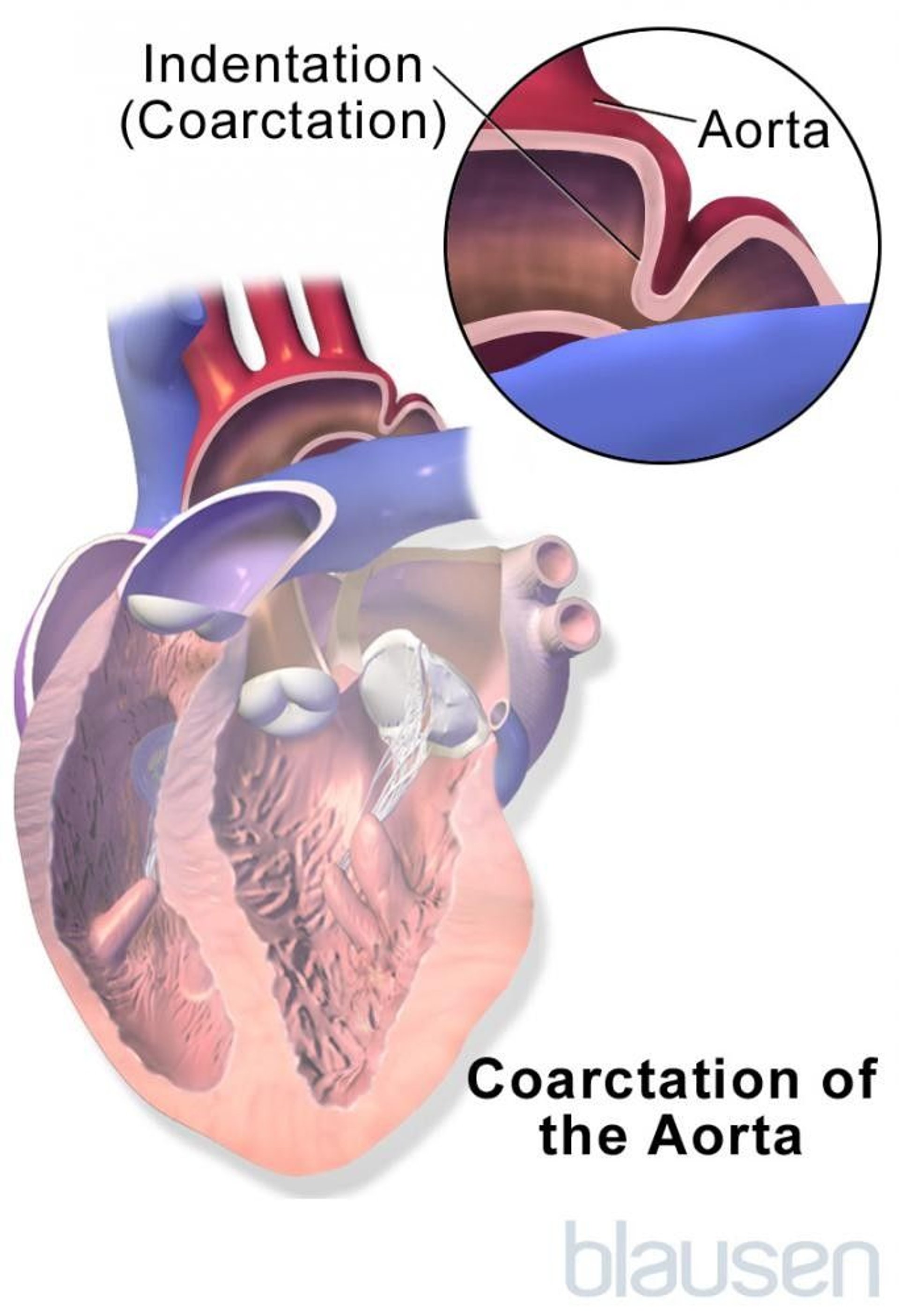
Coarctation of the aorta is a narrowing of part of the aorta, the main blood vessel bringing red oxygenated blood from the heart to the body.
The aorta narrows, causing the heart to pump harder to push blood through the narrowed aorta to get adequate blood flow to the lower half of the body.
Infants with severe coarctation become suddenly ill when they are a few days old, showing signs of heart failure and decreased blood flow to the lower body. Older children with coarctation often have no symptoms.
The diagnosis is suspected based on symptoms and findings during a doctor's examination and is confirmed with echocardiography.
Most children with coarctation need corrective surgery or balloon angioplasty to widen the narrowed aorta.
(See also Overview of Heart Defects.)
Coarctation, a heart birth defect, is narrowing of just one segment of the aorta, not the whole aorta. The narrowing is typically located across from the point where the ductus arteriosus joins the aorta. The ductus arteriosus is a blood vessel connecting the two great arteries leaving the heart, the pulmonary artery and the aorta (see Normal Fetal Circulation). In the womb and during the first few days of life, the ductus is open, so blood flowing through it bypasses the narrowed section of aorta.
Coarctation may reduce blood flow to the lower half of the body, including the kidneys, liver, and other organs in the abdomen. The blood pressure is lower than normal in the legs and tends to be higher than normal in the arms. Coarctation is a serious but treatable cause of high blood pressure. A heart murmur (a sound created by turbulent blood flow through narrowed or leaking heart valves or through abnormal heart structures) is sometimes present.

Without treatment, severe coarctation can be fatal in newborns. Less severe coarctation eventually strains and enlarges the heart and causes high blood pressure. Coarctation makes the child susceptible to heart infection (infective endocarditis) and bleeding in the brain. Children with coarctation often have other heart defects, such as aortic valve stenosis or an atrial or a ventricular septal defect.
Symptoms of Coarctation of the Aorta
Infants with mild coarctation usually have few or no symptoms. Infants with moderate coarctation may have rapid, sometimes labored breathing, a fast heart beat, poor feeding, grayish skin coloration, decreased number of wet diapers, and irritability or lethargy.
With a severe coarctation in infancy, blood can flow to the lower portion of the aorta (at a point past its narrowing) through the open connection between the aorta and the pulmonary artery, the ductus arteriosus. Symptoms usually do not occur until the ductus closes, usually when the newborn is a few days old. After the closure, the blood supplied through the ductus disappears, sometimes causing sudden loss of almost the entire blood supply to the lower body. Sudden, catastrophic heart failure and low blood pressure can result.
Most older children with coarctation do not have symptoms. Occasionally, children with coarctation have headaches or nosebleeds because of high blood pressure in the upper body or leg pains during exercise because the blood supply to the legs is insufficient.
Diagnosis of Coarctation of the Aorta
Echocardiography
Sometimes computed tomography (CT) or magnetic resonance imaging (MRI)
Coarctation is usually suspected when a doctor notices a heart murmur or differences in pulses or blood pressures between the arms and legs when doing a physical examination. A heart murmur is a sound created by turbulent blood flow through narrowed or leaking heart valves or through abnormal heart structures. In addition, the blood pressure measured in the arms may be quite high.
Echocardiography (ultrasonography of the heart) and, sometimes, CT or MRI confirms the diagnosis.
Electrocardiography (ECG) and chest x-rays are typically done. They may show an enlarged heart.
Treatment of Coarctation of the Aorta
Sometimes a medication, a prostaglandin
Surgery, balloon angioplasty, or stent placement
Treatment depends on the severity of the coarctation and the symptoms it causes.
Infants with severe symptoms due to coarctation require emergency treatment, including
Treatment with a prostaglandin, a medication that can reopen the ductus arteriosus to improve blood flow to the lower half of the body
Other medications to strengthen the heart's pumping
Emergency surgery to widen the narrowing
Surgery is the preferred treatment for moderate or severe coarctation in infants and small children. The narrow portion of the aorta may be cut out and the two normal sections then sewn together, or a patch can be used to enlarge the narrow portion of the aorta. Sometimes, tissue from the blood vessel supplying the left arm (the subclavian artery) is used to create this patch.
A very mild coarctation that causes no symptoms may not get detected early in life. It may get noticed when children are somewhat older and the aorta has become narrower. A doctor may notice stronger pulses and a higher blood pressure in the arm than the leg. In such children, surgery and balloon angioplasty are options for repairing the narrowed aorta. Balloon angioplasty is done during cardiac catheterization, in which a thin tube (catheter) with a balloon at its tip is passed through a blood vessel in the arm or leg into the narrowed aorta. The balloon is inflated, widening the narrowed aorta. Sometimes, an expandable flexible tube (stent) is inserted to help keep the aorta from narrowing again.
Some children who had surgery for coarctation develop scar tissue in the aorta at the site of repair. Such scar tissue narrows the aorta and develops most commonly in infants who needed emergency surgery very early in life. Balloon angioplasty with or without stent placement is usually a very effective treatment for this recurrent narrowing.
Children need to take antibiotics before visits to the dentist and before certain surgeries (such as on the respiratory tract) for the 6-month period following repair. These antibiotics are used to prevent a serious heart infection called endocarditis.
More Information
The following English-language resources may be useful. Please note that THE MANUAL is not responsible for the content of these resources.
American Heart Association: Common Heart Defects: Provides an overview of common birth defects of the heart for parents and caregivers
American Heart Association: Infective Endocarditis: Provides an overview of infective endocarditis, including summarizing antibiotic use, for parents and caregivers


