


Ultrasonography is a type of medical imaging that uses high-frequency (ultrasound) waves to produce a moving image of internal organs and other tissues. Echocardiography is ultrasonography of the heart.
Ultrasonography of the heart (echocardiography) is one of the most widely used procedures for diagnosing heart disorders because it provides excellent images and is
Noninvasive
Harmless
Relatively inexpensive
Widely available
Also, because ultrasonography does not use x-rays, it does not cause any radiation exposure.
Ultrasonography is also used in the diagnosis of disorders affecting blood vessels in other parts of the body.
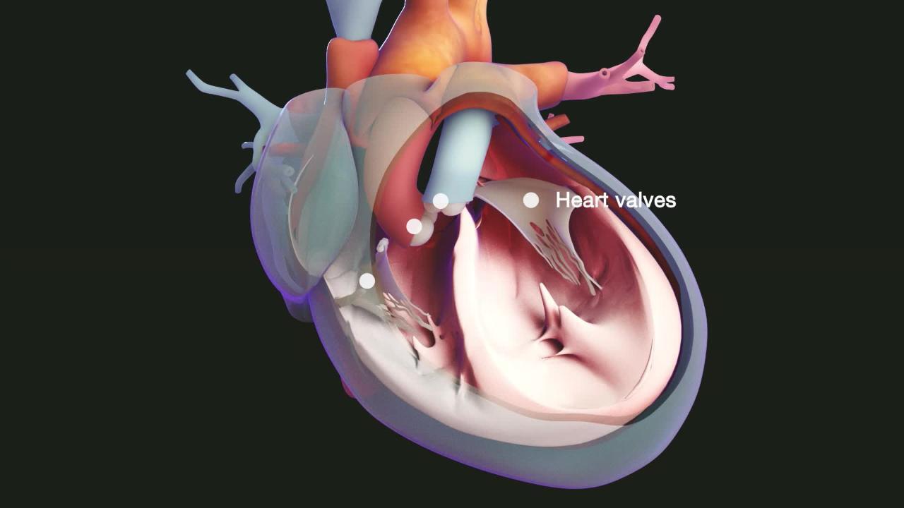
Echocardiography can be used to detect whether the heart muscle is moving normally and how much blood the heart is pumping out with each beat. This procedure can also detect abnormalities in the heart's structure, such as defective heart valves, birth defects (such as holes in the walls between the heart's chambers), and enlargement of the heart's walls or chambers, as occurs in people with high blood pressure, heart failure, or impairment of the heart's muscular walls (cardiomyopathy).

Echocardiography can also be used to detect pericardial effusion, in which fluid accumulates between the two layers of the sac that envelops the heart (pericardium), and constrictive pericarditis, in which scar tissue forms throughout the pericardium. It also can detect dissection of the aorta, a tearing within the layers of the wall of the aorta.
Sometimes echocardiography is done as part of a stress test.

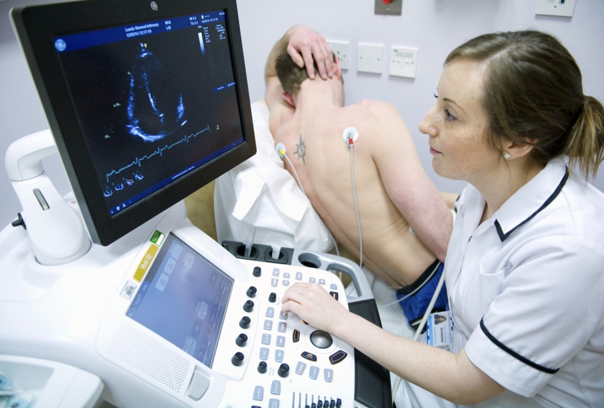
LTH NHS TRUST/SCIENCE PHOTO LIBRARY
The main types of ultrasonography are
Two-dimensional
Three-dimensional
Doppler
Color Doppler
Strain imaging
Two-dimensional ultrasonography, the most widely used technique, produces realistic two-dimensional images in computer-generated "slices." "Stacking" the slices together can re-create a three-dimensional structure.
Doppler ultrasonography shows the direction and velocity of blood flow and thus can detect turbulent flow due to narrowing or blockage of blood vessels.
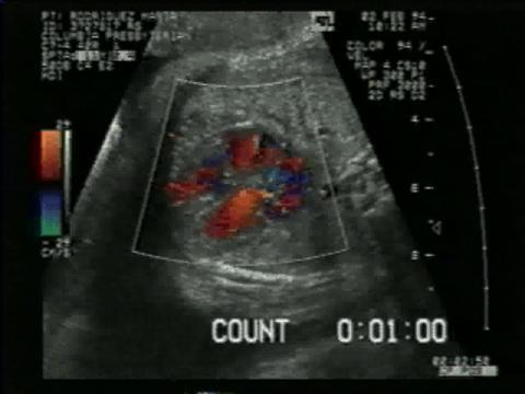
Color Doppler ultrasonography shows the different directions of blood flow in different colors.
Doppler ultrasonography and color Doppler ultrasonography are commonly used to help diagnose disorders affecting the heart and the arteries and veins in the trunk, legs, and arms. Because these procedures can show the direction and rate of blood flow in the chambers and blood vessels of the heart, they enable doctors to evaluate the structure and function of these parts. For example, doctors can determine if the heart valves open and close properly, if and how much they leak when closed, and if blood flows normally. Abnormal connections between an artery and a vein or between heart chambers can also be detected.
Strain imaging is an echocardiographic technique that is increasingly used. It measures changes in the motion of the heart muscle. Strain imaging can diagnose heart disease before changes are visible with conventional echocardiography, differentiate between different causes of heart disease, and predict prognosis in a variety of heart diseases, including heart failure.
How echocardiography is done
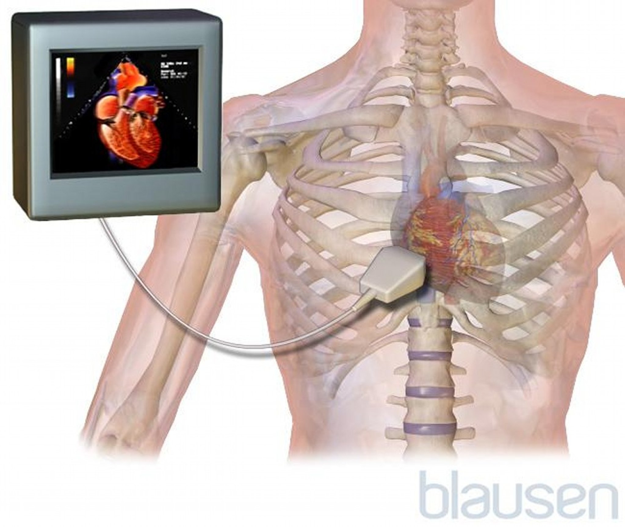
The ultrasound waves are emitted by a probe that both emits and detects ultrasound waves (transducer). The transducer can be placed
On the person's chest (transthoracic)
In the person's esophagus (transesophageal)
Sometimes on a catheter inside the heart (intracardiac)
In transthoracic echocardiography (the most common type), the transducer is handheld and placed on the chest over the heart. The examiner places gel on the chest under the transducer to help transmit the sound waves into the chest. The transducer is connected to a computer that displays an image on a monitor and stores the image digitally. By varying the placement and angle of the transducer, doctors can view the heart and nearby major blood vessels from various angles and thus get an accurate picture of heart structure and function. During various portions of the examination, people will need to hold their breath for about 10 seconds to ensure clear images are obtained. Transthoracic echocardiography is painless and takes 20 to 30 minutes.
Sometimes doctors use a portable ultrasonography machine at the person's bedside in order to determine specific information quickly. Often portable technology is used when people are receiving care in an emergency department or intensive care unit.

Transesophageal echocardiography can be used if doctors need to obtain greater clarity or to analyze the aorta or structures at the back of the heart (particularly the left atrium or left ventricle). For this procedure, a small flexible tube with an ultrasound transducer at the tip is passed down the person's throat into the esophagus so that the transducer lies just behind the heart. Because this procedure is uncomfortable, the person is sedated and the throat is numbed with an anesthetic spray. Transesophageal echocardiography is also used when regular echocardiography is difficult to do because of obesity, lung disorders, or other technical problems or when doctors are looking for specific diseases, such as endocarditis of the mitral valve or aortic valve or a clot within the heart.
Intracardiac echocardiography is a rare type of echocardiography that is done when a person is having a procedure on the heart such as a procedure to repair an atrial septal defect (hole in the heart). For intracardiac echocardiography, a small flexible tube with an ultrasound transducer at the tip is passed from a blood vessel in the groin directly into a chamber of the heart. The person having this procedure is usually sedated. Intracardiac echocardiography is used when doctors need to obtain detailed pictures of the heart that cannot be obtained using transthoracic or transesophageal echocardiography.


