


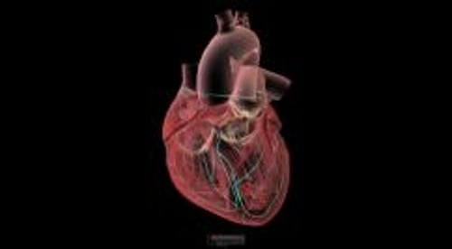
Electrocardiography (ECG) is a quick, simple, painless medical test that measures the heart’s electrical impulses.
During an ECG, the heart's electrical impulses are measured, amplified, and recorded. This record, the electrocardiogram (also known as an ECG), provides information about the
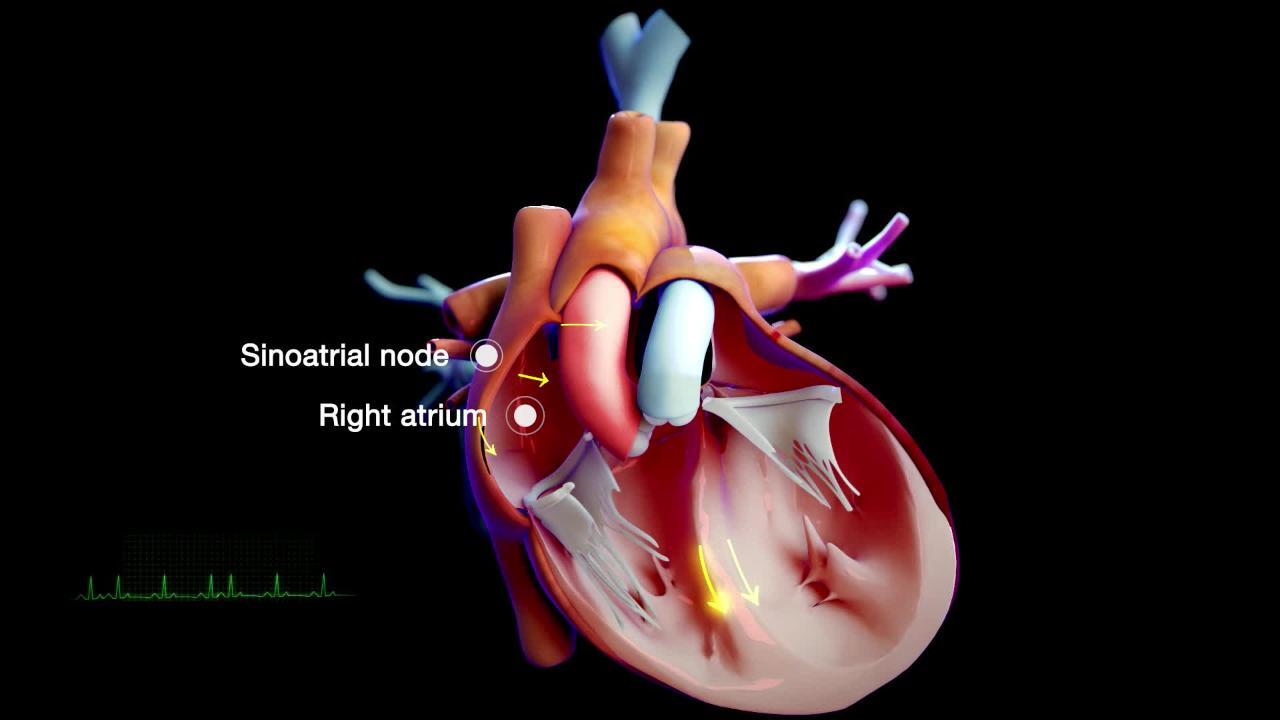
Part of the heart that triggers each heartbeat (the pacemaker, called the sinoatrial or sinus node)
Nerve conduction pathways of the heart
Rate and rhythm of the heart
Sometimes, the ECG can show that the heart is enlarged (for example, possibly due to high blood pressure) or that the heart is not receiving enough oxygen due to a blockage in one of the blood vessels that supply the heart (the coronary arteries).


From D Dubin, Rapid Interpretation of EKGs, 1996, Cover Inc., p. 95; with permission.
Usually, an ECG is obtained if a heart disorder is suspected. It is sometimes also obtained as part of a routine physical examination for middle-aged and older adults, even if they have no evidence of a heart disorder. It can be used as a basis of comparison with later ECGs if a heart disorder develops.
Abnormal heart rhythms and inadequate blood flow to the heart muscle may occur only briefly or unpredictably. To detect such problems, doctors may use continuous ambulatory electrocardiography, in which the ECG is recorded while the person engages in normal daily activities.
How ECG is done
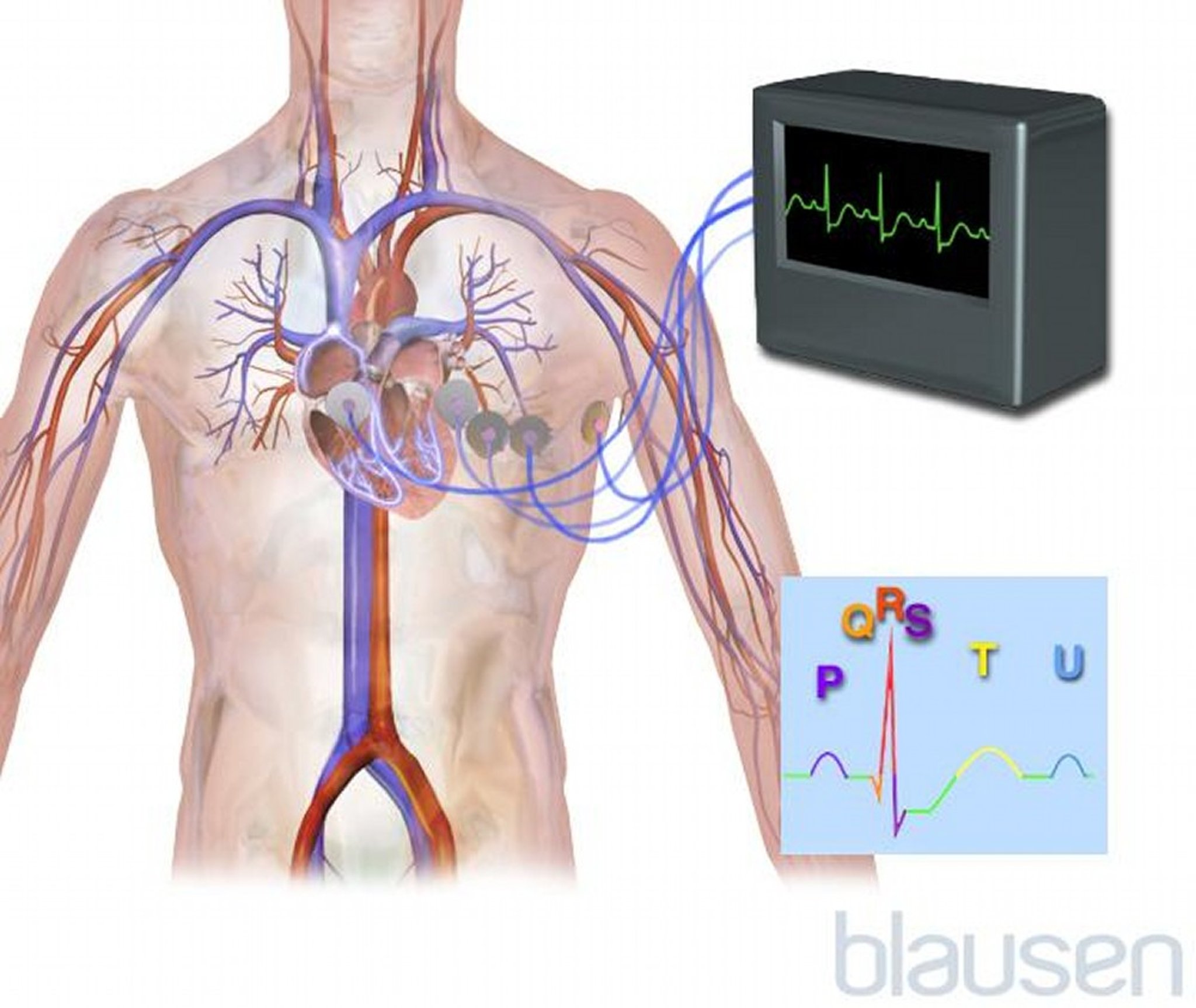
To obtain an ECG, an examiner places electrodes (small round sensors that stick to the skin) on the person's arms, legs, and chest. A standard ECG uses 12 electrodes (called a 12-lead ECG). These electrodes do not contain needles and are painless. If thick hair is present, the areas to which the electrodes are applied may first be shaved. These electrodes measure the magnitude and direction of electrical currents in the heart during each heartbeat. The electrodes are connected by wire s to a machine, which produces a record (tracing) for each electrode. Each tracing shows the electrical activity of the heart from different angles. The tracings constitute the ECG. ECG takes about 3 minutes and has no risks.
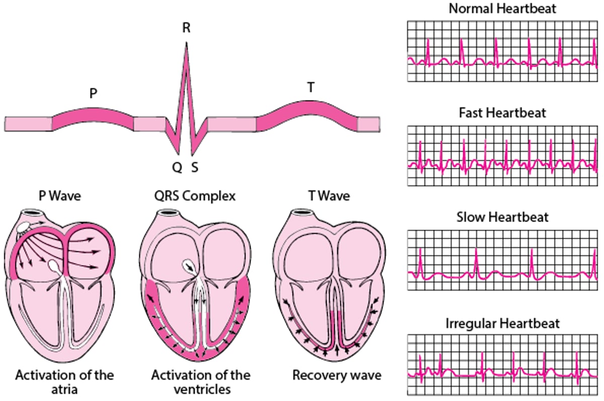
ECG: Reading the Waves
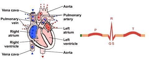
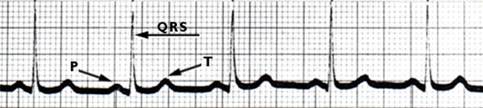
An electrocardiogram (ECG) represents the electrical current moving through the heart during a heartbeat. The current's movement is divided into parts, and each part is given an alphabetic designation in the ECG. Each heartbeat begins with an impulse from the heart's pacemaker (sinus or sinoatrial node). This impulse activates the upper chambers of the heart (atria). The P wave represents activation of the atria. Next, the electrical current flows down to the lower chambers of the heart (ventricles). The QRS complex represents activation of the ventricles. The electrical current then spreads back over the ventricles in the opposite direction. This activity is called the recovery wave, which is represented by the T wave. Many kinds of abnormalities can often be seen on an ECG. They include a previous heart attack (myocardial infarction), an abnormal heart rhythm (arrhythmia), an inadequate supply of blood and oxygen to the heart (ischemia), and excessive thickening (hypertrophy) of the heart's muscular walls. Certain abnormalities seen on an ECG can also suggest bulges (aneurysms) that develop in weak areas of the heart's walls. Aneurysms may result from a heart attack. If the rhythm is abnormal (too fast, too slow, or irregular), the ECG may also indicate where in the heart the abnormal rhythm starts. Such information helps doctors begin to determine the cause and the most appropriate treatment. |

Smartwatches
ECG technology is also available in smartwatches. Such smartwatches use a single electrode sensor placed on the wrist to measure the heart's electrical activity. This kind of ECG can detect arrhythmias, including atrial fibrillation (Afib), which is a type of abnormal heart rhythm. Unlike a standard 12-lead ECG, smartwatch ECGs allow for continuous monitoring of heart activity, instead of a single brief period. However, smartwatch ECGs are typically not as accurate as 12-lead ECGs and do not detect as many different cardiac conditions.


