Bronchoscopy is a direct visual examination of the voice box (larynx) and airways through a viewing tube (a bronchoscope).
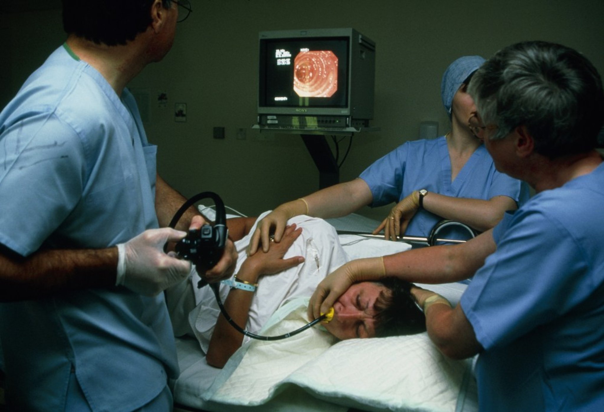
A bronchoscope, a thin viewing tube with a light, has a camera at the end that allows a doctor to look down through the larger airways (bronchi) into the lungs. Doctors can also pass small tools through the bronchoscope to allow them to take samples of lung or airway tissue to help diagnose lung disorders and to treat some lung disorders. Doctors pass the bronchoscope through the person's nose or mouth, down the windpipe, and into the airways.
Bronchoscopes may be
Flexible
Rigid
Most bronchoscopy procedures, particularly those used for diagnosis, are done using a flexible bronchoscope.
Some diagnostic and therapeutic procedures require the use of a rigid bronchoscope and are done under general anesthesia in a hospital. For example, removing a foreign object, controlling bleeding, or widening an airway may be best done through a rigid metal bronchoscope in an operating room.
Most procedures done with a flexible bronchoscope can be done in an outpatient setting, meaning the person is not admitted to the hospital. Sometimes the person is sedated before the procedure, and a topical (nasal and/or inhaled) anaesthetic is used.
(See also Medical History and Physical Examination for Lung Disorders and Overview of the Respiratory System.)
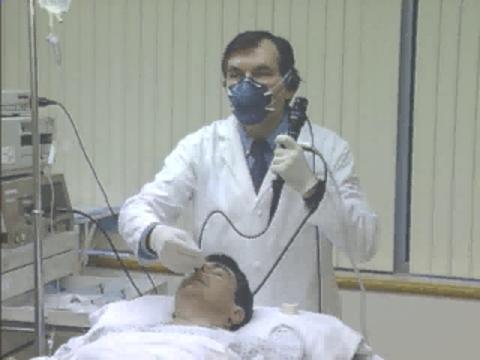
Understanding Flexible Bronchoscopy
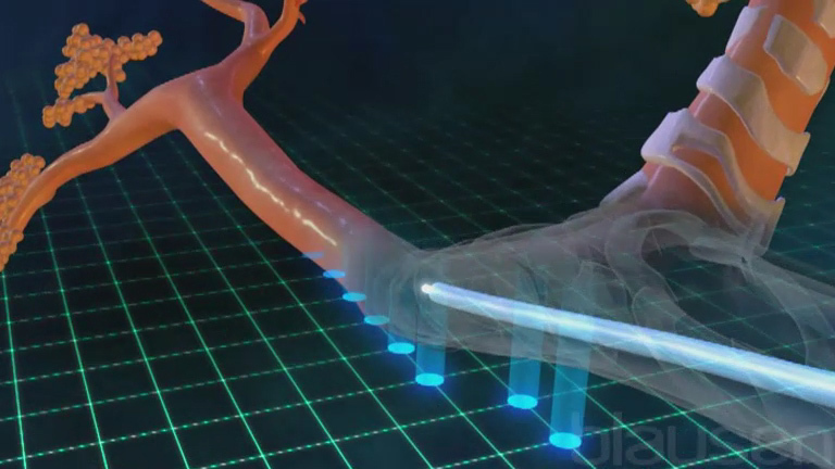
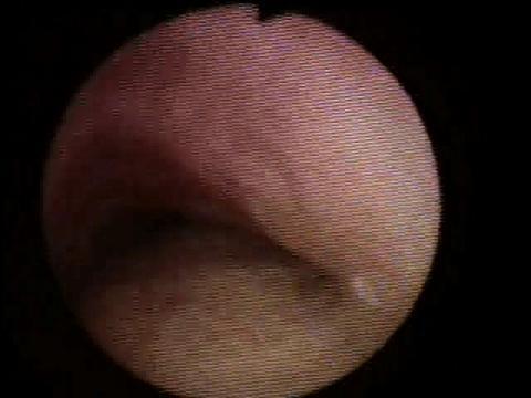
To view the airways directly, a doctor passes a flexible bronchoscope through a person's nostril or into the mouth and down into the airways. The circular inset shows the doctor's view. |

A bronchoscope can be used to
Assess the airways and voice box (larynx) for injury in people who have been burned or may have inhaled smoke
Determine the cause of lung infections (such as pneumonia) if there is concern that the cause is unusual bacteria or may be difficult to treat (for example, in people who have AIDS or other immune system deficiencies)
Examine airways and take tissue specimens from areas that may be cancerous
Investigate a source of bleeding in the lungs

ANTONIA REEVE/SCIENCE PHOTO LIBRARY
Bronchoscopy can help doctors treat certain conditions. For example, the bronchoscope can be used to
Be a guide over which a tube can be inserted to assist breathing (tracheal intubation)
Place medications or substances in specific areas of the lung
Remove secretions, blood, pus, and foreign bodies
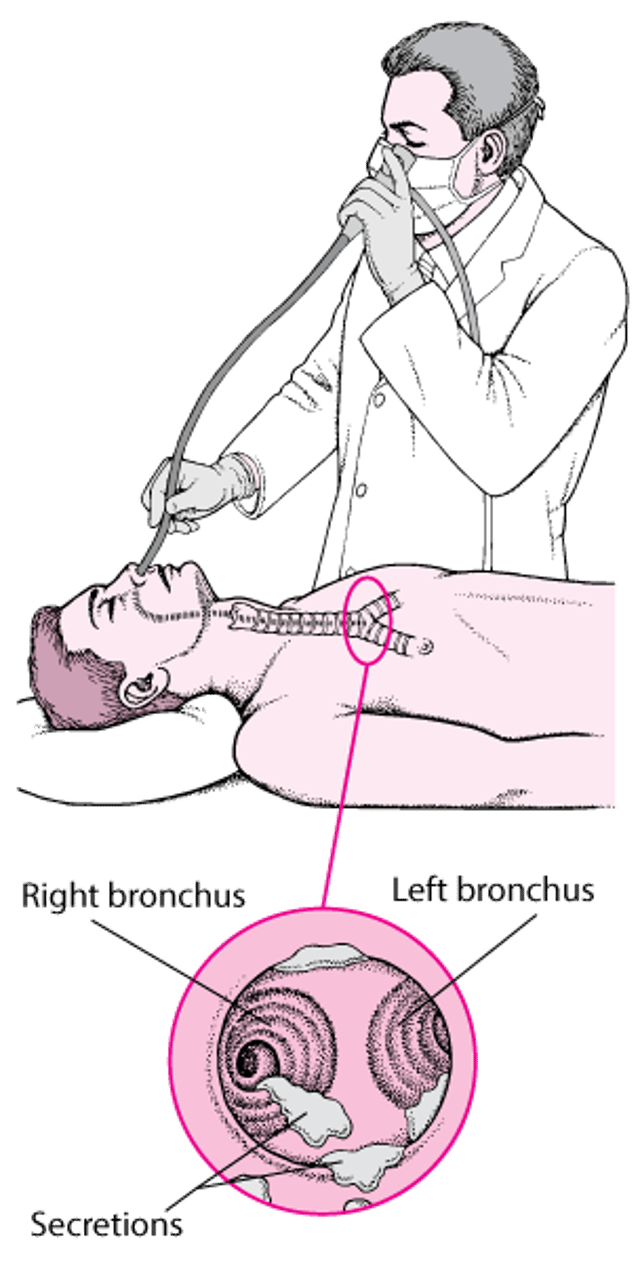
For at least 6 hours before bronchoscopy, the person should not eat or drink. Sedation is often given to people having flexible bronchoscopy, and general anesthesia is usually given to those undergoing rigid bronchoscopy. In flexible bronchoscopy, the throat and nasal passage are sprayed with an anesthetic, and the bronchoscope is passed through a nostril, mouth, or breathing tube and into the airways of the lungs.
After bronchoscopy, the person is observed for 2 to 4 hours. If a tissue specimen was removed, a chest x-ray may be taken to check for complications, such as bleeding or pneumothorax (air in the chest but outside the lungs).
Procedures Done With Bronchoscopy
Sometimes, as part of a bronchoscope examination, doctors do additional procedures to collect specimens for examination in a laboratory.
Bronchoalveolar lavage is a procedure doctors can use to collect specimens from the smaller airways and air sacs (alveoli) that cannot be seen through the bronchoscope. After wedging the bronchoscope into a small airway, a doctor administers salt water (saline) through the instrument. The fluid is then suctioned back into the bronchoscope, bringing cells and any bacteria with it. Examination of the material under the microscope helps in diagnosing infections and cancers. The fluid can also be placed into containers containing special nutrients and left alone for a period of time to see whether bacteria grow (culturing), which is a better way to diagnose infections.
Transbronchial lung biopsy involves obtaining a specimen (pieces) of lung tissue by using forceps passed through a channel in a bronchoscope. The bronchoscope is threaded into progressively smaller airways until it reaches the area of concern. A doctor may use a fluoroscope (an imaging device that uses x-rays to show internal body structures on a screen) for guidance in identifying the area of concern. Such guidance can also decrease the risk of accidentally perforating the lung and causing leakage of air into the pleural space (pneumothorax). Although transbronchial lung biopsy increases the risk of complications during bronchoscopy, it provides additional diagnostic information and may make major surgery unnecessary.
Transbronchial needle aspiration is sometimes done. In this procedure, a needle is passed through the bronchoscope into the bronchial wall. The needle may be passed through the wall of a large airway under direct visualization or through the wall of a small airway using an x-ray machine for visualization. A doctor may be able to extract cells from suspicious lymph nodes to examine under a microscope. Endobronchial ultrasonography (EBUS) can be used to help guide the needle biopsy.
With bronchial washing, a salt water solution is injected through the bronchoscope and subsequently aspirated from the airways. Cells and other materials retrieved can then be examined to help identify lung disease. Bronchial washing is similar to bronchoalveolar lavage but uses much less water.
In bronchial brushing, a brush is advanced through the bronchoscope and used to loosen cells from areas of concern so they can be examined.
Navigational bronchoscopy uses software to combine images obtained from computed tomography (CT) to construct a "virtual" map (in 3 dimensions) of the lung. The map is used to identify a path to an area targeted for biopsy but that is deep (in the periphery) in the lung. Such areas are more difficult to access using bronchoscopy than areas that are not as far in the lung periphery. During navigational bronchoscopy, images from the bronchoscope can be combined with the virtual map in real time to help guide the bronchoscope along the best path. Electromagnetic navigational bronchoscopy is a form of navigational bronchoscopy in which images derived from an electromagnetic field are also used.