


Mitral stenosis is narrowing of the mitral orifice that impedes blood flow from the left atrium to the left ventricle. The usual cause is rheumatic fever. Common complications are pulmonary hypertension, atrial fibrillation, and thromboembolism. Symptoms are those of heart failure; signs include an opening snap and a diastolic murmur. Diagnosis is by physical examination and echocardiography. Prognosis is good. Medical treatment includes diuretics, beta-blockers or rate-limiting calcium channel blockers, and anticoagulants. Effective treatment for more severe disease consists of balloon commissurotomy, surgical commissurotomy, or valve replacement.
(See also Overview of Cardiac Valvular Disorders.)
In mitral stenosis, mitral valve leaflets become thickened and immobile and the mitral orifice becomes narrowed due to fusion of the commissures and the presence of shortened, thickened, and matted chordae tendineae.
The most common cause is
However, many patients do not recall having had rheumatic fever. In regions with high prevalence of rheumatic fever, patients often present by age 30 years, usually with commissural fusion of pliable, noncalcified leaflets. In regions with low prevalence, patients typically present between ages 50 and 70 years and with calcified fibrotic leaflets with subvalvular involvement in addition to commissural fusion.
An increasingly prevalent cause in older patients is mitral annular calcification with extension of calcification into the leaflets, causing them to stiffen and not open fully.
Women comprise 80% of rheumatic fever and mitral annular calcification cases.
Occasionally, mitral stenosis is congenital or occurs after radiation therapy.
If the valve cannot close completely, mitral regurgitation (MR) may coexist with mitral stenosis. Patients with mitral stenosis due to rheumatic fever may also have lesions of the aortic or tricuspid valve or both.
Left atrial (LA) size and pressure increase progressively to compensate for mitral stenosis; pulmonary venous and capillary pressures also increase and may cause secondary pulmonary hypertension, leading to right ventricular (RV) heart failure, tricuspid regurgitation, and pulmonic regurgitation. Rate of progression varies.
LA enlargement predisposes to atrial fibrillation (AFib), a risk factor for thromboembolism. The faster heart rate and loss of atrial contraction with onset of atrial fibrillation often lead to sudden worsening of symptoms.
Symptoms and Signs of Mitral Stenosis
Symptoms of mitral stenosis correlate poorly with disease severity because the disease often progresses slowly, and patients unconsciously reduce their activity. Many patients are asymptomatic until they become pregnant or atrial fibrillation develops.
Initial symptoms are usually those of heart failure (eg, exertional dyspnea, orthopnea, paroxysmal nocturnal dyspnea, fatigue).
Symptoms typically do not appear until 15 to 40 years after an episode of rheumatic fever. In medically underserved regions, young children may become symptomatic because streptococcal infections are not treated with antibiotics, and recurrent infections are common.
Paroxysmal or chronic atrial fibrillation further reduces blood flow into the left ventricle (LV), precipitating pulmonary edema and acute dyspnea when ventricular rate is poorly controlled. Atrial fibrillation may also cause palpitations. In up to 15% of patients not taking anticoagulants, it causes systemic embolism with symptoms of stroke or other organ ischemia.
Less common symptoms include hemoptysis due to rupture of small pulmonary vessels and pulmonary edema, particularly during pregnancy when blood volume increases. Hoarseness due to compression of the left recurrent laryngeal nerve by a dilated LA or pulmonary artery (Ortner syndrome) and symptoms of pulmonary hypertension and RV failure may also occur.
Mitral stenosis may cause signs of cor pulmonale. The classic facial appearance in mitral stenosis, a plum-colored malar flush, occurs only when cardiac output is low and pulmonary hypertension is severe; cause is cutaneous vasodilation and chronic hypoxemia. In patients with dark skin, the malar flush may produce a darker shade of the patient's skin.
Occasionally, the initial symptoms and signs of mitral stenosis are those of an embolic event such as stroke. Endocarditis is rare in mitral stenosis unless mitral regurgitation is also present.
Palpation
Palpable first and second heart sounds (S1 and S2) may be detected. S1 is best palpated at the apex, and S2 is best felt at the upper left sternal border. The pulmonic component of S2 (P2) is responsible for the impulse and results from pulmonary hypertension.
An RV impulse (heave) palpable at the left sternal border may accompany jugular venous distention when pulmonary hypertension is present and RV diastolic dysfunction develops.
Auscultation
Loud S1
Early diastolic opening snap
Low-pitched decrescendo-crescendo rumbling diastolic murmur
Auscultatory findings in mitral stenosis include a loud S1 caused by the leaflets of a stenotic mitral valve closing abruptly (M1); it is heard best at the apex. S1 may be absent when the valve is heavily calcified and immobile. A normally split S2 with an exaggerated P2 due to pulmonary hypertension is also heard (see table Distinguishing the Murmurs of Tricuspid Stenosis and Mitral Stenosis).
Most prominent is an early diastolic opening snap as the leaflets billow into the LV, which is loudest close to left lower sternal border; it is followed by a low-pitched decrescendo-crescendo rumbling diastolic murmur, heard best with the bell of the stethoscope at the apex (or over the palpable apex beat) at end-expiration when the patient is in the left lateral decubitus position. The opening snap may be soft or absent if the mitral valve is calcified; the snap moves closer to S2 (increasing duration of the murmur) as mitral stenosis becomes more severe and LA pressure increases.
The diastolic murmur increases after a Valsalva maneuver (when blood pours into the LA), after exercise, and in response to maneuvers that increase afterload (eg, squatting, isometric handgrip). The murmur may be softer or absent when an enlarged RV displaces the LV posteriorly and when other disorders (pulmonary hypertension, right-sided valve abnormalities, atrial fibrillation with fast ventricular rate) decrease blood flow across the mitral valve. The presystolic crescendo is caused by increased flow with atrial contraction. However, the closing mitral valve leaflets during LV contraction may also contribute to this finding but only at the end of short diastoles when LA pressure is still high.
Diastolic murmurs that may coexist with the MS murmur are
Early diastolic murmur of coexisting aortic regurgitation (AR), which may be conducted to the apex
Diastolic flow murmur in the presence of severe MR
Mid-diastolic murmur of obstructing left atrial myxoma or ball thrombus (rare)
Graham Steell murmur (a soft decrescendo diastolic murmur heard best along the left sternal border and caused by pulmonic regurgitation secondary to severe pulmonary hypertension)
Diagnosis of Mitral Stenosis
Echocardiography
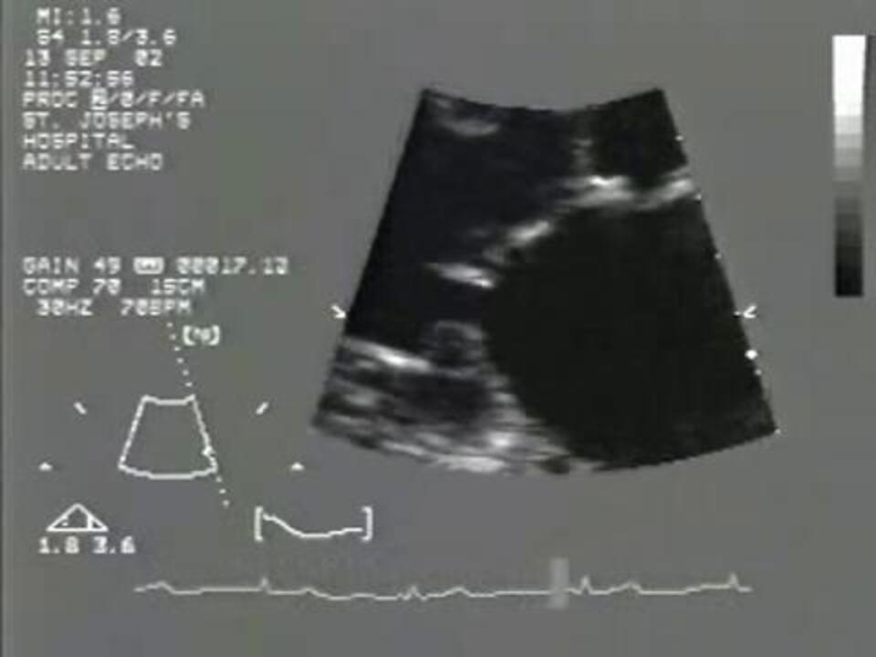
Diagnosis of mitral stenosis is suspected clinically and confirmed by echocardiography. Typically, 2-dimensional echocardiography shows abnormal valve and subvalvular structures. It also provides information about the degree of valvular calcification and stenosis and LA size. Doppler echocardiography provides information about the transvalvular gradient and pulmonary artery pressure. The normal area of the mitral valve orifice is 4 to 5 cm2.
Severity of mitral stenosis is characterized echocardiographically as
Moderate: Valve area > 1.5 to 2.5 cm2 or diastolic pressure half-time < 150 msec
Severe: Valve area ≤ 1.5 cm2 or diastolic pressure half-time ≥ 150 msec; symptoms are often present
However, the relationship between the area of the valve orifice and symptoms is not always consistent. Color Doppler echocardiography detects associated mitral regurgitation. Transesophageal echocardiography (TEE) can be used to detect or exclude small LA thrombi, especially those in the LA appendage, which usually cannot be seen transthoracically. TEE also can better assess mitral regurgitation when mitral calcification causes acoustic shadowing of the left atrium from the transthoracic window.
An ECG and chest x-ray are usually obtained.
The ECG may show LA enlargement, manifest as a P wave lasting > 0.12 msec with prominent negative deflection of its terminal component (duration: > 0.04 msec; amplitude: > 0.10 mV) in V1; broad, notched P waves in lead II; or both. Right axis QRS deviation and tall R waves in V1 suggest RV hypertrophy.
Chest x-ray usually shows straightening of the left cardiac border due to a dilated LA appendage, and widening of the carina. With barium in the esophagus, the lateral chest x-ray will show the dilated LA displacing the esophagus posteriorly. The main pulmonary artery (trunk) may be prominent; the descending right pulmonary artery diameter is ≥ 16 mm if pulmonary hypertension is significant. The upper lobe pulmonary veins may be dilated. A double shadow of an enlarged LA may be seen along the right cardiac border. Horizontal lines in the lower posterior lung fields (Kerley B lines) indicate interstitial edema associated with high LA pressure.
Exercise testing helps quantify symptoms. Further information can be obtained from stress echocardiography evaluation of changes in valve gradient and pulmonary pressure.
Cardiac catheterization, indicated only for perioperative assessment of coronary artery disease (CAD) before surgical repair, can confirm elevated LA and pulmonary artery pressures, mitral gradient, and valve area.
Treatment of Mitral Stenosis
Diuretics and sometimes beta-blockers or calcium channel blockers
Sometimes anticoagulation for atrial fibrillation
Commissurotomy or valve replacement
Asymptomatic patients with mitral stenosis require no treatment other than appropriate prophylaxis against rheumatic fever recurrence. Surveillance with serial TTE is important, because RV enlargement and rise in RV systolic pressure can occur without patients noticing a change in functional state and without a decrease in mitral valve area. Early intervention may relieve pulmonary hypertension before it becomes permanent.
Mildly symptomatic patients usually respond to diuretics and, if sinus tachycardia or atrial fibrillation is present, to beta-blockers or calcium channel blockers, which can control ventricular rate.
Anticoagulation with a vitamin K antagonist (not a direct-acting oral anticoagulant [DOAC]) is indicated to prevent thromboembolism if patients have or have had atrial fibrillation, embolism, or a left atrial clot. Extended restoration of sinus rhythm is rarely possible. Anticoagulation may be considered in the presence of dense spontaneous contrast or an enlarged left atrium (M-mode diameter > 50 mm) because these findings are associated with thromboembolism, although anticoagulation for these patients has not been studied. All patients should be encouraged to continue at least low levels of physical exercise despite exertional dyspnea.
Antibiotic prophylaxis against endocarditis is no longer recommended except for patients who have had valve replacement (see table Recommended Endocarditis Prophylaxis During Oral-Dental or Respiratory Tract Procedures).
Timing of intervention
Moderate mitral stenosis intervention may be indicated when cardiac surgery is required for other indications. Patients who are symptomatic and have exercise-induced mean transmitral gradient > 15 mm Hg or pulmonary capillary occlusion pressure > 25 mm Hg may be considered for percutaneous balloon commissurotomy.
Severe mitral stenosis intervention is indicated when any symptoms are present if the valve is suitable for percutaneous balloon commissurotomy (may be considered in asymptomatic patients if pulmonary pressure is > 50 mm Hg or there is new-onset atrial fibrillation). Cardiac surgery is pursued only when symptoms are severe and for patients who are not candidates for percutaneous balloon commissurotomy, require other cardiac operations, or do not have access to the percutaneous procedure.
Choice of intervention
Percutaneous balloon commissurotomy is the procedure of choice for younger patients and for patients without heavily calcified valve commissures, subvalvular distortion, LA thrombi, or moderate or severe MR (see Table Grading of Mitral Regurgitation —1). In this fluoroscopic- and echocardiographic-guided procedure, a transvenous catheter with an inflatable distal balloon is passed transseptally from the right atrium to the LA and inflated to separate fused mitral valve commissures. Outcomes are equivalent to those of more invasive procedures. Complications are uncommon but include MR, embolism, and tamponade. About 75% of the atrial septal defects from the procedure close spontaneously and most of the rest have only clinically trivial left to right shunts, but percutaneous closure is occasionally necessary.
Surgical commissurotomy may be used in patients with severe subvalvular disease, valvular calcification, or LA thrombi. In this procedure, fused mitral valve leaflets are separated using a dilator passed through the left ventricle (closed commissurotomy) via a thoracotomy, or by direct vision (open commissurotomy) via a sternotomy. Choice of procedure is based on the surgeon’s experience and the morphology of the valve, although closed commissurotomy is done less frequently. Because of its greater risks, surgery is usually deferred until symptoms reach New York Heart Association class III (see table Classification of Heart Failure). During surgery, some clinicians ligate the left atrial appendage to reduce risk of thromboembolism.
Valve replacementwarfarin for 3 to 6 months postoperatively (see also Anticoagulation for patients with a prosthetic cardiac valve). Direct-acting oral anticoagulants (DOAC) are ineffective and should not be used.
When the etiology is annular calcification, there is no benefit from percutaneous balloon commissurotomy because there is no commissural fusion. Furthermore, surgical valve replacement is technically demanding because of the annular calcification and often high risk because many patients are older and have comorbidities. Therefore, intervention is delayed until symptoms become severe despite use of diuretic and rate control medications. Preliminary experience in patients who are not candidates for surgery suggests benefit from implantation of a transcatheter aortic valve replacement (TAVR) bioprosthesis in the mitral position.
Treatment reference
1. Otto CM, Nishimura RA, Bonow RO, et al: 2020 ACC/AHA Guideline for the Management of Patients With Valvular Heart Disease: Executive Summary: A Report of the American College of Cardiology/American Heart Association Joint Committee on Clinical Practice Guidelines. Circulation 143(5):e35–e71, 2021. doi: 10.1161/CIR.0000000000000932
Prognosis for Mitral Stenosis
The natural history of mitral stenosis varies, but the interval between onset of symptoms and severe disability is about 7 to 9 years. Outcome is affected by the patient’s preprocedural age and functional status, presence of pulmonary hypertension, and degree of mitral regurgitation. Symptomatic results of balloon or surgical commissurotomy are equivalent in patients with valves that are not calcified. However, after a variable period of time, function deteriorates in most patients due to restenosis, and valve replacement may become necessary. Risk factors for death are atrial fibrillation and pulmonary hypertension. Cause of death is most commonly heart failure or pulmonary or cerebrovascular embolism.
Key Points
Mitral stenosis is usually caused by rheumatic fever, although calcific degeneration is increasingly observed in older patients.
Pulmonary hypertension and atrial fibrillation may develop.
The risk of thromboembolism in patients with atrial fibrillation and mitral stenosis is very high and is treated with a vitamin K antagonist, not a direct-acting oral anticoagulant.
Heart sounds include a loud first heart sound and an early diastolic opening snap followed by a low-pitched decrescendo-crescendo rumbling diastolic murmur, heard best at the apex at end-expiration when the patient is in the left lateral decubitus position; the murmur increases after a Valsalva maneuver, exercise, squatting, and isometric handgrip.
Mildly symptomatic patients usually respond to diuretics and, if sinus tachycardia or atrial fibrillation is present, to beta-blockers or calcium channel blockers for rate control.
Severely symptomatic patients and those with evidence of pulmonary hypertension require commissurotomy or valve replacement.


