


Caries is tooth decay, commonly called cavities. The symptoms—tender, painful teeth—appear late. Diagnosis is based on inspection, probing of the enamel surface with a fine metal instrument, and dental x-rays. Treatment involves removing affected tooth structure and restoring it with various materials. Fluoride, diligent dental hygiene, sealants, and proper diet can prevent virtually all caries.
Etiology of Caries
Caries is caused by acids produced by bacteria in dental plaque. Plaque is, at first, a soft, thin film of bacteria, mucin, dead epithelial cells, and food debris that develops on the tooth surface within about 24 hours after the tooth is cleaned. Streptococcus mutans species are a group of related bacteria that grow in plaque and can cause caries. Some strains are more cariogenic than others. Eventually (commonly, after 72 hours), soft plaque mineralizes, mainly with calcium, phosphate, and other minerals, becoming calculus (hard plaque or tartar), which cannot easily be removed with a toothbrush.
Risk factors
There are several risk factors for caries:
Inadequate plaque control
Dental defects
Diet, particularly frequent consumption of dietary carbohydrates and sugars
High-acid and/or low-fluoride environment
Characteristics of saliva, including reduced salivary flow (eg, due to drugs, radiation therapy, systemic disorders that cause salivary gland dysfunction), and buffering capacity and pH
Hereditary genetic factors
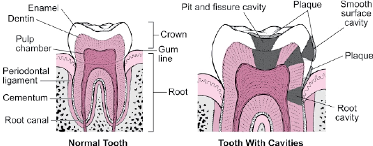
Many teeth have open enamel pits, fissures, and grooves, which may extend from the surface to the dentin (see figure Types of cavities). These defects may be wide enough to harbor bacteria but too narrow to clean effectively. They predispose teeth to caries.
Frequent dietary exposure to carbohydrates and sugars promotes the growth of plaque-forming bacteria. Development of severe early childhood caries (rampant caries) in deciduous teeth suggests prolonged contact with infant formula, milk, or juice, typically when an infant goes to bed with a bottle (baby or nursing bottle caries). Thus, bedtime bottles should contain only water.
A tooth surface is more susceptible to caries when it is poorly calcified, has low fluoride exposure, and/or is in an acidic environment. Typically, decalcification begins when the pH at the tooth falls below 5.5 (eg, when lactic acid–producing bacteria colonize the area or when people drink soft drinks, sports drinks or energy drinks, which commonly have pH values below 5.5).

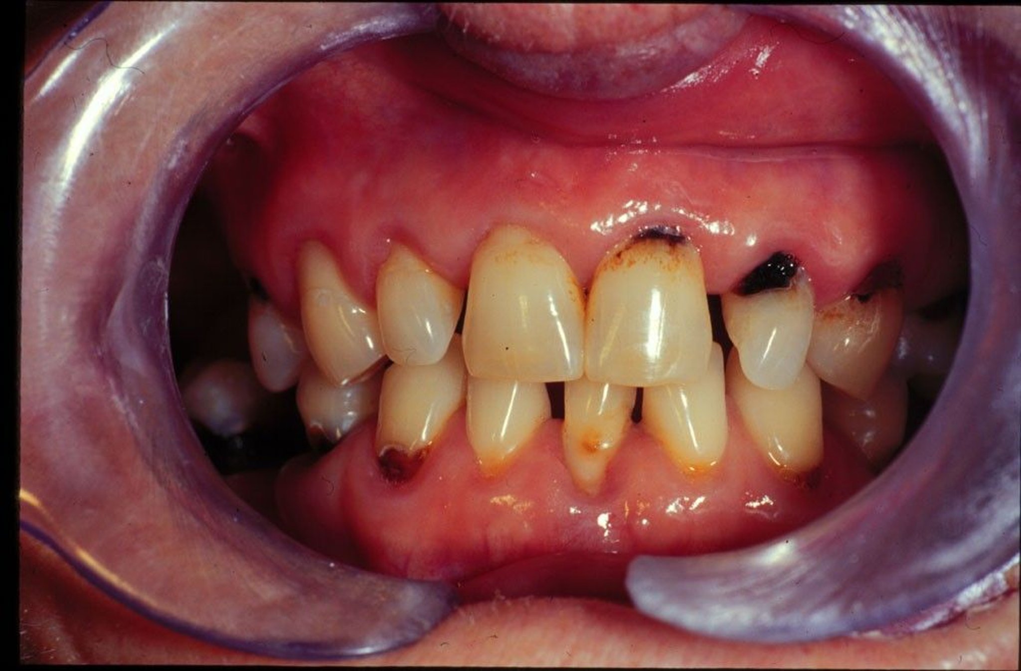
Image provided by Jonathan A. Ship, DMD.
Older patients often take drugs that reduce salivary flow, predisposing to caries. They also have a higher incidence of root caries because of gingival recession, exposure of root surfaces, and declining manual dexterity (causing ineffective oral hygiene).
Types of cavities

Complications
Untreated caries leads to tooth destruction, infections, and the need for extractions and replacement prostheses. Premature loss of deciduous teeth may shift the adjacent teeth, hindering eruption of their permanent successors.
General reference
Wong A, Subar PE, Young DA: Dental caries: An update on dental trends and therapy. Adv Pediatr 64(1):307-330, 2017. doi: 10.1016/j.yapd.2017.03.011
Symptoms and Signs of Caries
Caries initially involves only the enamel and causes no symptoms. Caries that invades the dentin causes pain, first when hot, cold, or sweet foods or beverages contact the involved tooth, and later with chewing or percussion. Pain can be intense and persistent when the pulp is severely involved (pulpitis).
Diagnosis of Caries
Direct inspection
Sometimes use of x-rays or special testing instruments

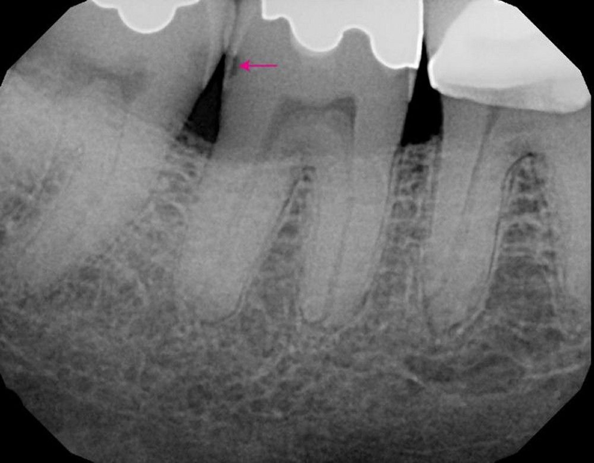
Image provided by James Ubertalli, DMD.
Routine, frequent clinical evaluation (every 3 to 12 months depending on a patient's caries risk as assessed by a dentist) identifies early caries at a time when minimal intervention prevents its progression. A thin probe, sometimes special dyes, and transillumination by fiberoptic lights are used, although infrequently these are supplemented by new devices that detect caries by changes in electrical conductivity, laser reflectivity, or transillumination (including use of near-infrared light devices). However, x-rays are still most important for detecting caries, determining the depth of involvement, and identifying caries under existing restorations.
Treatment of Caries
Restorative therapy
Sometimes a root canal and crown
Remineralization of teeth
Remineralization of teeth with incipient caries (which is confined to the enamel) is usually attempted with improved home care (brushing and flossing), cleanings, prescriptions for high-fluoride toothpastes, and multiple fluoride applications at the dental office. Other topical products promoted for remineralization contain calcium and phosphates but are less effective in remineralizing caries than fluoride. Silver diammine fluoride (SDF), commercially available in the United States since 2015, can arrest and remineralize carious lesions. However, because SDF permanently stains caries black, it is applied primarily on deciduous teeth.
Restoration of teeth
The primary treatment of caries that has entered dentin is removal by drilling, followed by filling of the resultant defect.
Fillings for occlusal surfaces of posterior teeth, which bear the brunt of mastication, must be composed of strong materials, including
Silver amalgam
Composite resins
Silver amalgam combines silver, mercury, copper, tin, and occasionally zinc, palladium, or indium. Amalgam is inexpensive and lasts an average of 14 years. However, if oral hygiene is good and if amalgam was placed using a rubber dam for isolation from saliva, many amalgam fillings last > 40 years. Amalgam is a highly durable material with annual failure rates lower than that of composite resins; therefore, amalgam restorations last longer and are more resistant to secondary caries than composite restorations (1).
Amalgam use is decreasing for 2 main reasons:
The results are not as aesthetically pleasing as those achieved with either composites or glass ionomers.
There are environmental concerns about the removal and disposal of the mercury content of amalgam.
Although concern has been raised about "mercury poisoning," the number of amalgam fillings a person has bears no relationship to their blood mercury levels. Replacing amalgam is not recommended because it is expensive, damages tooth structure, actually increases patient exposure to mercury, and requires the use of amalgam separators to prevent disposal of its mercury content into the environment. Because of recent environmental concerns about the disposal of the mercury content of traditional fillings, there is a trend toward using dental materials other than amalgam.
Composite resins, which have a more acceptable appearance, have long been used in anterior teeth, where aesthetics are primary and the forces of chewing are minimal. Many patients request them in posterior teeth as well, and they are now commonly used there. Early-generation composite resins under high occlusal stress generally lasted less than half as long as amalgam and tended to develop recurrent decay because the composite resin shrinks when it hardens and expands and contracts with heat and cold more than the tooth or other filling materials. The current generation of composites more closely simulates enamel hardness, does not appear to have the same incidence of recurrent caries as earlier materials, and may also last longer. Use of bonded composite resin restorations allows for more conservation of tooth structure compared to amalgam preparations.
Glass ionomer, a tooth-colored filling, releases fluoride when in place, a benefit for patients especially prone to tooth decay. It is also used to restore areas damaged by overzealous brushing. Glass ionomer is not as aesthetic as composite and it should not be used on chewing surfaces because it has a high wear rate. Resin-modified glass ionomer materials are also available and provide some improved aesthetics compared to conventional glass ionomers.
If decay leaves too little dentin to retain a full or partial coverage restoration, a dentist replaces the missing dentin with cement, amalgam, composite, or other materials. Sometimes a post must be inserted into one or more roots to support a gold, silver, or composite core, which replaces the coronal dentin. This procedure necessitates a root canal filling, in which an opening is made in the tooth and the pulp is removed. The root canal system is thoroughly debrided, shaped, and then filled with gutta-percha. The outer tooth surfaces (what would have been the enamel) are then reduced so that an artificial crown, usually made of metal, porcelain, or ceramic can be placed. Crowns for anterior teeth are made of, or covered with, porcelain or ceramic.
Treatment reference
1. Moraschini V, Fai CK, Alto RM, et al: Amalgam and resin composite longevity of posterior restorations: A systematic review and meta-analysis. J Dent 43(9):1043-1050, 2015. doi: 10.1016/j.jdent.2015.06.005
Prevention of Caries
Regular brushing and flossing
Fluoride in water, toothpaste, or both
Regular professional cleanings
For most people, caries is preventable. Cavities first form on permanent teeth in the early teens to late 20s. Caries-prone people typically have low exposure to fluoride and a relatively cariogenic microflora acquired from their mothers and through social contact.

Maintaining good oral hygiene is especially important, as are dietary factors, particularly minimizing intake of sugar and acidic drinks and eating a diet rich in sources of calcium.
Removal of plaque at least every 24 hours, usually by brushing and flossing, helps prevent dental caries. The gingival third of the tooth is the most important area to clean but is the area most often neglected. Brushing with an electric toothbrush for 2 minutes is excellent; brushing with a manual soft toothbrush for 3 to 4 minutes suffices. Using excess toothpaste, particularly an abrasive type, may erode the teeth. Dental floss is placed between each of the teeth, curved against the side of each tooth, and moved up and down 3 times, going just beneath the gingival margin. Flosses that are very thin or coated with wax or polytetraethylene can be used for exceptionally tight contacts between teeth or rough filling margins.
Teeth with fluoride incorporated into their enamel are more resistant to acidic decalcification and more readily recalcify when pH increases. If drinking water is not adequately fluoridated, oral fluoride supplements are recommended for children from 6 months through 16 years. Fluoride supplements in pregnant women have not been shown to prevent caries in offspring and thus are no longer recommended (1).
Oral supplements for children are available in toothpastes, liquids (for use in a dropper for infants), gels, and tablets. Tablets should be swished in the mouth to enhance topical fluoride absorption before swallowing. The dose must be selected according to the amount of fluoride present in the drinking water and diet, the age of the child, and whether topical fluoride is being used in toothpaste and/or applied during dental care. Excessive fluoride exposure may cause dental fluorosis , causing mild to severe discoloration of teeth. Also, fluoridated toothpaste should be used by people of all ages. Because young children may swallow toothpaste when brushing, which can lead to fluorosis, they should use children's toothpastes, which contain lower amounts of fluoride.
Fluoridation offers less protection against caries in pits and fissures than against those on smooth surfaces. Deep, narrow pits and fissures require use of sealants (resin materials that adhere tightly to the surface of the enamel) to prevent nutrients from reaching bacteria, reducing their growth and acid production.
If these measures do not decrease cavity formation, more intensive therapy is aimed at changing the flora. After cavities are treated, pits and fissures, which can harbor S. mutans,S. mutans. To encourage this repopulation, xylitol
xylitol, as mentioned above, from the time of the child’s birth to the age at which the mother no longer samples the child’s food (a hypothesized mode of transfer). Prenatal interventions may reduce transfer of cariogenic bacteria from mother to child and delay caries formation in the child.
For prevention of caries in deciduous teeth (once they have erupted) in infants, bedtime bottles should contain only water.
Prevention reference
1. Richards D: No evidence that fluoride supplements taken during pregnancy prevent caries. Evid Based Dent 19(3):73, 2018. doi: 10.1038/sj.ebd.6401320
Key Points
Caries is caused by acids produced by bacteria in dental plaque.
Risk factors include preexisting tooth defects, low saliva flow, an acidic oral environment, frequent exposure to carbohydrates and sugar in the diet, and inadequate exposure to fluoride.
Treatment of incipient caries confined to the enamel may be attempted with remineralization through improved home care (brushing and flossing), cleanings, prescriptions for high-fluoride toothpastes, and multiple fluoride applications at the dental office.
Treatments of caries that have entered the dentin involve drilling out the decayed area and restoring the defect with amalgam, composite resin, or glass ionomer.
Prevention involves meticulous regular brushing, flossing, and professional cleaning; adequate fluoride must be available in toothpaste and, when not present in drinking water, as oral supplements for children.


