


Atopic dermatitis is a chronic relapsing inflammatory skin disorder with a complex pathogenesis involving genetic susceptibility, immunologic and epidermal barrier dysfunction, and environmental factors. Pruritus is a primary symptom; skin lesions range from mild erythema to severe lichenification to erythroderma. Diagnosis is by history and examination. Treatments include counseling on appropriate skin care, avoidance of triggers, and topical corticosteroids and immunosuppressants. Control of pruritus and superinfections is also important. Severe cases may require systemic immunosuppressive treatment. Childhood atopic dermatitis frequently resolves or lessens significantly by adulthood.
(See also Definition of Dermatitis.)
Etiology of Atopic Dermatitis
Atopic dermatitis primarily affects children in urban areas or high-income countries, and prevalence has increased over the last 30 years; up to 20% of children and 10% of adults in high-income countries are affected. Most people with the disorder develop it before age 5, many of them before age 1; however, atopic dermatitis may even begin during late adulthood. The unproven hygiene hypothesis suggests that decreased early childhood exposure to infectious agents (ie, because of more rigorous hygiene regimens at home) may increase the development of atopic disorders and autoimmunity to self-proteins.
Many patients or their family members who have atopic dermatitis also have allergic asthma and/or immediate-type hypersensitivities that manifest, for example, as allergic seasonal or perennial rhinoconjunctivitis. The triad of atopic dermatitis, allergic rhinoconjunctivitis, and asthma is called atopy or atopic diathesis. Other dermatologic features of atopy include xerosis, ichthyosis/palmar hyperlinearity (ie, more prominent skin lines on the palms), keratosis pilaris, an infraorbital skinfold (Dennie-Morgan fold), thinning of the lateral eyebrows (Hertoghe sign), wool intolerance (irritation and itching triggered by skin contact with wool), white dermographism (vasoconstriction, causing whitening of the skin in response to scratching), and increased transepidermal water loss (in nonaffected as well as affected skin).
Pathophysiology of Atopic Dermatitis
All of the following contribute to the development of atopic dermatitis:
Genetic factors
Epidermal barrier dysfunction
Immunologic mechanisms
Environmental triggers
Genes implicated in atopic dermatitis are those encoding epidermal and immunologic proteins. A major predisposing factor for atopic dermatitis is the existence in many patients of a loss-of-function mutation in the gene encoding for the filaggrin protein (1). Filaggrin is a component of the cornified cell envelope produced by differentiating keratinocytes. It is ultimately critical to building the hygroscopic barrier of the stratum corneum (also called the natural moisturizing factor). About 10% of European populations are heterozygous carriers of loss-of-function filaggrin mutations. Presence of these mutations (as well as more intragenic copy mutations) increases the risk of more severe atopic dermatitis and higher IgE levels. Filaggrin mutations are also associated with peanut allergy and asthma, even in the absence of atopic dermatitis.
These recent molecular insights provide new understanding of atopic dermatitis and how the cutaneous inflammation, a T-cell–mediated delayed-type hypersensitivity, relates to allergic conditions with immediate-type hypersensitivities such as asthma and allergic rhinitis (hay fever). The epidermal skin barrier defect due to filaggrin mutations explains the development of xerosis and the predisposition to skin irritation. This results in the manifestation of atopic dermatitis, which is not an allergic reaction. The cutaneous inflammation, in contrast, is a T-cell–mediated delayed-type hypersensitivity and includes a Th2-dominant component in the skin. This hypersensitivity downregulates antimicrobial peptides (eg, beta-defensins), which explains why patients with atopic dermatitis are predisposed to developing bacterial and viral skin infections. The increased penetration of skin irritants and allergens also results in a Th2-dominant inflammation, which also drives IgE production and predisposes to immediate-type hypersensitivities. However, because the major mechanism mediating atopic dermatitis is delayed cell-mediated immunity, avoidance of immediate-type allergens (eg, pollen, dust mites) usually does not improve atopic dermatitis. Although immediate-type hypersensitivities (eg, asthma, allergic rhinitis) result from the skin barrier defect, they do not drive the T-cell–mediated cutaneous inflammation of atopic dermatitis.
In patients with atopic diathesis, atopic dermatitis typically precedes allergic rhinoconjunctivitis and asthma. Sometimes, this sequence is called the "atopic march" and occurs because the skin barrier defect is the primary deficiency in atopic conditions.
Pathophysiology reference
1. Brown SJ, McLean WH: One remarkable molecule: Filaggrin. J Invest Dermatol 132(3 Pt 2):751–762, 2012. doi: 10.1038/jid.2011.393
Symptoms and Signs of Atopic Dermatitis
Atopic dermatitis usually appears in infancy, as early as 3 months of age.
In the acute phase, lesions are intensely pruritic, red, thickened, scaly patches or plaques that may become eroded due to scratching.
In the chronic phase, scratching and rubbing create skin lesions that appear dry and lichenified.
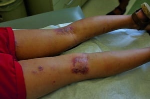
Distribution of lesions is age specific. In infants, lesions characteristically occur on the face, scalp, neck, eyelids, and extensor surfaces of the extremities. In older children and adults, lesions occur on flexural surfaces such as the neck and the antecubital and popliteal fossae.
Intense pruritus is a key feature. Itch often precedes lesions and worsens with dry air, sweating, local irritation, wool garments, and emotional stress.
Common environmental triggers of symptoms include
Excessive bathing or washing
Harsh soaps
Staphylococcus aureus skin colonization
Sweating
Rough fabrics and wool
© Springer Science+Business Media

Image provided by Thomas Habif, MD.

Image provided by Thomas Habif, MD.
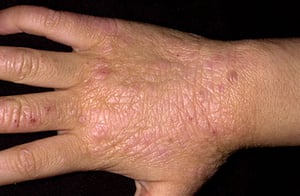
Photo provided by Thomas Habif, MD.


© Springer Science+Business Media

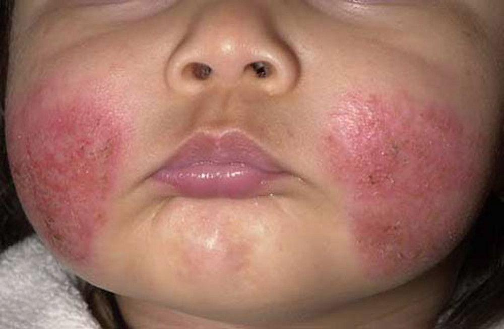
Image provided by Thomas Habif, MD.

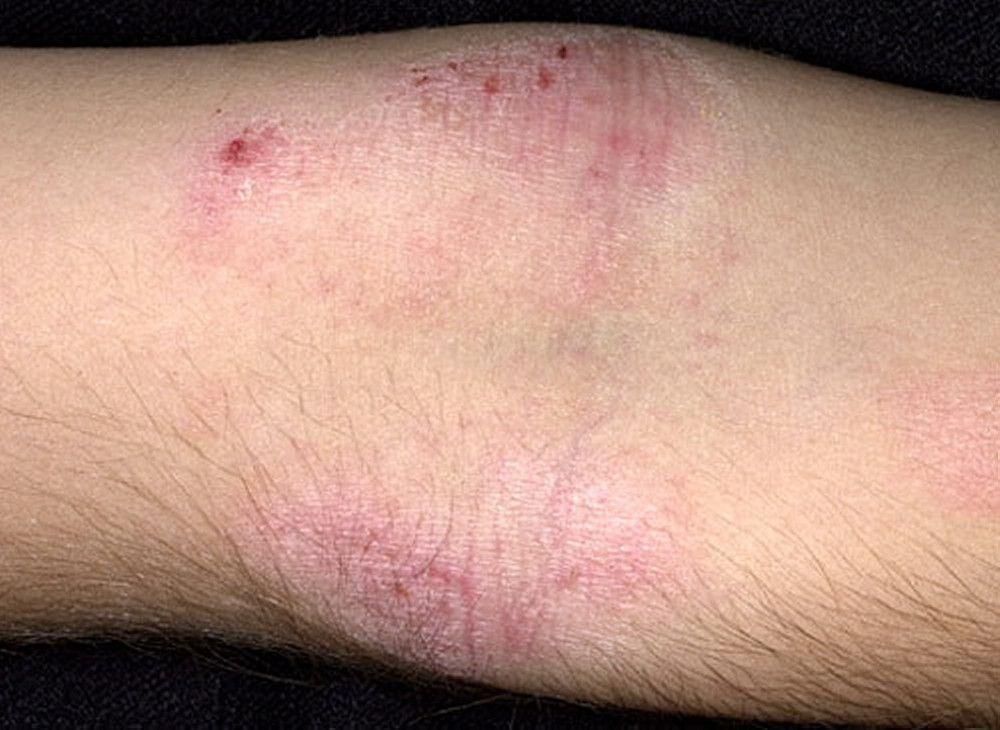
Image provided by Thomas Habif, MD.


Photo provided by Thomas Habif, MD.
Complications of atopic dermatitis
Secondary bacterial infections (superinfections), especially staphylococcal and streptococcal infections (eg, impetigo, cellulitis) are common.
Erythroderma (erythema that covers more than 70% of the body surface area) is rare but can result when atopic dermatitis is severe.

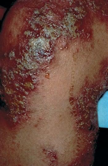
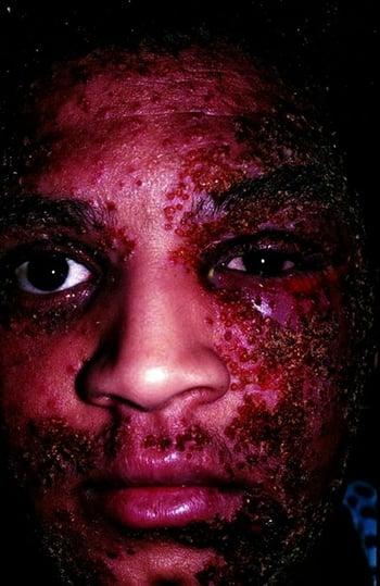
Eczema herpeticum is an infection with herpes simplex virus (HSV) of the skin that is more diffuse and widespread than those in nonatopic patients. Eczema herpeticum is commonly the primary HSV infection, but can also result from recurrent infection, in patients with atopic dermatitis. It manifests as grouped vesicles in areas of active or recent dermatitis, although normal skin can be involved. High fever and adenopathy may develop after several days. Occasionally, this infection can become systemic, which may be fatal. Sometimes the eye is involved, causing a painful corneal lesion. Eczema vaccinatum is a similar complication due to smallpox vaccination, in which the vaccinia virus disseminates and may become life-threatening. Atopic patients should therefore not receive smallpox vaccinations.

Atopic patients are also prone to other viral skin infections (eg, common warts, molluscum contagiosum).
Patients with atopic dermatitis also have a higher risk of allergic contact reactions. For example, contact allergies to nickel, the most common contact allergen, are twice as common as in nonatopic patients. Frequent use of topical products exposes patients to many potential allergens, and allergic contact dermatitis caused by these products may complicate the treatment of atopic dermatitis.
Diagnosis of Atopic Dermatitis
Clinical evaluation
Diagnosis of atopic dermatitis is clinical. History (eg, personal or family history of allergic rhinoconjunctivitis, seasonal or perennial; asthma) is helpful. See table Diagnostic Clinical Features in Atopic Dermatitis for the modified clinically relevant guidelines of care for the diagnosis and assessment of atopic dermatitis (2014) from the American Academy of Dermatology.
Atopic dermatitis is sometimes difficult to differentiate from other dermatoses. The diagnosis of atopic dermatitis depends on excluding other conditions, such as the following (1):
Contact dermatitis (irritant or allergic)
Photosensitivity dermatoses
Immune deficiency diseases
Erythroderma of other causes
Although a personal and/or family history of atopy and the distribution of lesions are helpful in making the diagnosis of atopic dermatits, the following distribution patterns can help with differentiation:
Psoriasis is typically easy to recognize by its sharply demarcated, thick, erythematous, and scaly plaques. It is usually extensoral rather than flexural and may include typical nail findings not common in atopic dermatitis (eg, oil spots, nail pits).
Seborrheic dermatitis most commonly affects the face (eg, nasolabial folds, eyebrows, glabellar region, scalp).
Nummular dermatitis is not flexural, and lichenification is rare. (However, coin-shaped, nummular plaques can occur in atopic dermatitis [nummular atopic dermatitis].)
Erythroderma caused by atopic dermatitis can be difficult to differentiate from erythroderma caused by other skin disorders.
Pearls & Pitfalls
|
There is no definitive laboratory test for atopic dermatitis. However, testing for type I allergies (prick, scratch, and intracutaneous testing or measurement of allergen-specific IgE levels and of total IgE levels) can help confirm atopic diathesis. Cultures for S. aureus are not done routinely but can help determine sensitivities to systemic antimicrobials when impetiginization is suspected (eg, with yellowish crusts).
Diagnosis reference
1. Eichenfield LF, Tom WL, Chamlin SL, et al: Guidelines of care for the management of atopic dermatitis: Section 1. Diagnosis and assessment of atopic dermatitis. J Am Acad Dermatol 70(2):338–351, 2014. doi: 10.1016/j.jaad.2013.10.010
Treatment of Atopic Dermatitis
Supportive care (including counseling on appropriate skin care and avoidance of precipitating factors)
Antipruritics
Topical corticosteroids
Topical calcineurin inhibitors
Topical Janus kinase (JAK) inhibitor (eg, ruxolitinib)
Phototherapy, particularly narrow-band ultraviolet B
Systemic immunosuppressants
Systemic biologic agents
Treatment of superinfections
Treatment of atopic dermatitis is most effective when addressing the underlying pathophysiologic processes.
Counseling on appropriate skin care and avoidance of triggers helps patients address the underlying skin barrier defect. Scratching of itchy lesions commonly increases itching and thus more scratching. Breaking this itch–scratch cycle is important.
Inflammatory flare-ups can be curtailed with topical immunosuppressants, phototherapy, and, if necessary, systemic immunosuppressants.
Almost all patients with atopic dermatitis can be treated as outpatients, but patients who have severe superinfections or erythroderma may need to be hospitalized.
(See also the American Academy of Dermatology’s management and treatment of atopic dermatitis with topical therapies [2014] and management and treatment with phototherapy and systemic agents [2014].)
Supportive care
General skin care should be focused on the most common sources of skin irritation—excessive washing as well as harsh soaps:
Limiting the frequency and length of washing and bathing (showers/baths should be limited to once daily; sponge baths can be substituted to decrease the number of days with full baths)
Limiting the temperature of bathing water to lukewarm
Avoiding excessive rubbing and instead patting skin dry after showering/bathing
Applying moisturizers (ointments or creams—ceramide-containing products are particularly useful)
For skin superinfection (eg, when yellowish crusting suggests impetigo), bathing with diluted bleach
Oral antihistamines< 12 years.
Reduction of emotional stress is useful and helps break the itch–scratch cycle. Stress can affect the family (eg, being kept awake by a crying baby) as well as the patient (eg, being unable to sleep because of itching).
Dietary changes intended to eliminate exposure to allergenic foods are generally unnecessary, are ineffective, and increase stress unnecessarily. Atopic dermatitis is driven by food allergies only very rarely.
Fingernails should be cut short to minimize excoriations and secondary infections.
Topical corticosteroids
Topical corticosteroids are the mainstay of therapy.
Creams or ointments applied 2 times a day are effective for most patients with mild or moderate disease. Low- to mid-potency corticosteroids often control skin inflammation, but resolution of chronically inflamed, lichenified skin typically requires high-potency corticosteroids. Dermatologic adverse effects of topical corticosteroids, such as thinning of the skin, striae distensae, and skin infections, may occur depending on the potency of the corticosteroid used, the duration of its use, and the skin location where it is used. Prolonged, widespread use of high-potency corticosteroids should be avoided, particularly in infants, to avoid adrenal suppression. Corticosteroids should always be stopped once the skin inflammation is controlled and should never be used to prevent recurrences.
Systemic corticosteroids typically provide good emergency relief, but long-term use should be avoided due their multiple adverse effects. Because atopic dermatitis tends to flare when systemic corticosteroids are stopped, other treatments are typically required to manage tapering and discontinuation of systemic corticosteroids.
Topical calcineurin inhibitors
Topical tacrolimus and pimecrolimus are calcineurin inhibitors. They are T-cell inhibitors and can be used for mild to moderate atopic dermatitis or when corticosteroid adverse effects are a concern.
Burning or stinging after application is usually transient and abates after a few days. Flushing is less common.
Topical crisaborole
Crisaborole is applied to areas of eczema 2 times a day. It cannot be used on mucous membranes.
Burning or stinging after application is the most common adverse effect.
Topical ruxolitinib
Ruxolitinib 1.5% cream can be used for the short-term and noncontinuous treatment of mild to moderate atopic dermatitis in immunocompetent patients 12 years of age and older whose disease is not adequately controlled with other topical prescription therapies or when those therapies are not advisable.
Ruxolitinib is applied as a thin layer 2 times a day to up to 20% of the body surface area for up to 8 weeks.
Phototherapy
Phototherapy with narrow-band ultraviolet B (UVB) is helpful for extensive atopic dermatitis, particularly when appropriate skin care and topical treatments fail to control inflammation. Because narrow-band UVB has much higher efficacy than the previously used broad-band UVB, psoralen plus UVA (PUVA) therapy is rarely used anymore. Phototherapy with narrow-band UVB has not been shown to increase skin cancer risk, but this remains a concern, particularly when used in children or for extended periods of time. This risk needs to be weighed against risks of other systemic treatments after failure of appropriate skin care and topical treatments.
If office-based phototherapy is not available or too inconvenient, home phototherapy is a good alternative. Several home phototherapy devices have programmable features that allow specialists to limit and supervise a patient's use of the device.
Natural sun exposure is an alternative when phototherapy is not available.
Systemic immunosuppressants
Risks include serious infections, death, cancer, cardiovascular events, and thrombosis.
Systemic biologic agents
Two targeted systemic immunosuppressants (biologics) are currently available for the treatment of moderate to severe atopic dermatitis.
Dupilumab is a fully human monoclonal IgG4 antibody that blocks the signaling of IL-4 and IL-13, both proinflammatory Th2 cytokines, in atopic dermatitis. It is available for the treatment of moderate to severe atopic dermatitis in patients 6 years of age or older and is recommended for patients whose disease is not adequately controlled with other treatments.
The most common adverse effects are injection site reactions, conjunctivitis, blepharitis, keratitis, eye pruritus, oral herpes, other herpes simplex virus infections, dry eye, and eosinophilia.
Tralokinumab-ldrm is a fully human monoclonal antibody against the IL-13 receptors alpha 1 and alpha 2. It is available for the treatment of moderate to severe atopic dermatitis in adults whose disease is not adequately controlled with topical therapies.
The most common adverse effects are upper respiratory infections, conjunctivitis, injection site reactions, and eosinophilia.
Treatment of superinfections
Antistaphylococcal antibiotics,impetigo, folliculitis, or furunculosis. Staphylococcus aureus is the most common bacterium causing skin infections in patients with atopic dermatitis and is often resistant to methicillin (methicillin-resistant S. aureus [MRSA]).
Staphylococcus aureus, a potential source of recurrent impetiginization.
Eczema herpeticum is treated with systemic antivirals (eg, acyclovir
Prognosis for Atopic Dermatitis
Atopic dermatitis in children often abates by age 5 years, although exacerbations are common throughout adolescence and into adulthood.
Girls and patients with severe disease, early age of onset, family history, and associated allergic rhinitis or asthma are more likely to have prolonged disease. Even in these patients, atopic dermatitis frequently resolves or lessens significantly by adulthood.
Atopic dermatitis may have long-term psychologic sequelae as children confront the challenges of living with a visible, sometimes disabling skin disease during their formative years.
Key Points
Atopic dermatitis is common, particularly in developed nations, affecting up to 20% of children and 10% of adults.
A genetically determined skin barrier defect predisposes to inflammation with skin irritants and thus atopic dermatitis.
Common triggers include excessive washing and bathing.
Common findings vary with age and include pruritus and scaly erythematous patches and plaques and lichenification in the antecubital and popliteal fossae and on the eyelids, neck, and wrists.
Superinfections (particularly S. aureus infections and eczema herpeticum) are common.
Atopic dermatitis frequently resolves or lessens significantly by adulthood.
First-line treatments include moisturizers, topical corticosteroids, and antihistamines as needed for pruritus.
For disease unresponsive to topical therapy, consider phototherapy or systemic immunosuppressants.
More Information
The following English-language resources may be useful. Please note that THE MANUAL is not responsible for the content of these resources.
American Academy of Dermatology (AAD): Section 1: Diagnosis and assessment of atopic dermatitis (2014)
AAD: Section 2: Management and treatment of atopic dermatitis with topical therapies (2014)
AAD: Section 3: Management and treatment with phototherapy and systemic agents (2014)


