


Contact dermatitis is inflammation of the skin caused by direct contact with irritants (irritant contact dermatitis) or allergens (allergic contact dermatitis). Symptoms include pruritus and sometimes a burning pain. Skin changes include erythema, scaling, skin swelling, and sometimes blistering and ulceration. The location depends on the site of contact. Diagnosis is by exposure history, examination, and sometimes skin patch testing. Treatment includes topical corticosteroids, antipruritics, and avoidance of irritants and allergens.
(See also Definition of Dermatitis.)
Pathophysiology of Contact Dermatitis
Contact dermatitis is caused by irritants or allergens.
Irritant contact dermatitis (ICD)
ICD is a nonspecific inflammatory reaction to toxic substances contacting the skin. Numerous substances can irritate the skin, including
Chemicals (eg, acids, alkalis, solvents, metal salts)
Soaps (eg, abrasives, detergents)
Plants (eg, poinsettias, peppers)
Chronic moisture (eg, from body fluids, urine, and saliva)
Properties of the irritant (eg, extreme pH, solubility in the lipid film on skin), environment (eg, low humidity, high temperature, high friction), and patient (eg, very young or old) influence the likelihood of developing ICD. ICD often can be divided into categories:
Acute ICD: Potent irritants, such as caustic chemicals, can damage the skin immediately, typically manifesting with acute burning or stinging pain.
Chronic or cumulative ICD: Less potent irritants require longer (chronic) or repeated (cumulative) periods of skin contact to cause ICD; these forms typically manifest with pruritus.
Occupational ICD is ICD caused by one or more of the many possible work-related skin irritants. It can be acute, chronic, or cumulative.
Atopic disorders increase risk of ICD because of impaired skin barrier function and lower threshold for skin irritation.
Phototoxic dermatitis ( see Chemical photosensitivity
Allergic contact dermatitis (ACD)
ACD is a type IV, T-cell–mediated, delayed-type hypersensitivity reaction to an environmental allergen that has 2 phases:
Sensitization to an antigen
Allergic response after reexposure
In the sensitization phase, allergens are captured by Langerhans cells (dendritic epidermal cells). When activated by innate immunity cascades, these cells migrate to regional lymph nodes, where they process and present the antigen to naive, antigen-specific T cells. When a naive T cell recognizes its antigen via binding to its T-cell receptor, it expands clonally and differentiates into memory/effector T cells. The sensitization phase, which is asymptomatic, may be brief (6 to 10 days for strong sensitizers such as poison ivy) or prolonged (years for weak sensitizers such as sunscreens, fragrances, and glucocorticoids). During differentiation, sensitized T cells become able to express cutaneous homing antigens (eg, cutaneous lymphocyte antigens) that enable them to migrate from cutaneous capillaries to the epidermis. When antigen-presenting cells present the antigen to the sensitized T cells, the T cells can expand and trigger an inflammatory reaction at that location (elicitation phase of ACD), resulting in the characteristic symptoms and signs of ACD.
Multiple allergens can cause ACD (see table Causes of Allergic Contact Dermatitis). Nickel sulfate is the most common contact allergen in most populations. The Toxicodendron species of plant (eg, poison ivy, poison oak, poison sumac) accounts for a large percentage of ACD, including moderate and severe cases. The offending allergen is urushiol.
Photoallergic contact dermatitis is a variant of ACD, in which a substance becomes sensitizing only after it undergoes structural change triggered by ultraviolet light. Reactions may extend to non–sun-exposed skin. Typical causes include fragrances (eg, musk ambrette, sandalwood), nonsteroidal anti-inflammatory drugs, and sunscreen filters.
Symptoms and Signs of Contact Dermatitis
Irritant contact dermatitis
Acute ICD is more painful than pruritic. Signs range from erythema, scaling, and edema to erosions, crusting, and blistering. Chronic and cumulative ICD is more often pruritic.
Allergic contact dermatitis
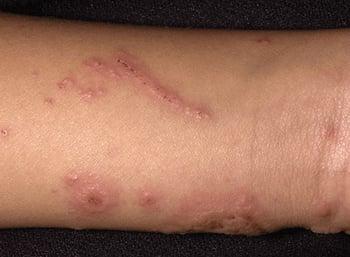
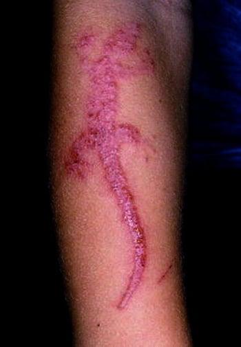
ACD is more pruritic than painful. Skin changes range from erythema, scaling, and edema, through vesiculation to severe swelling with bullae. Changes often occur in a pattern, distribution, or combination that suggests a specific exposure, such as linear streaking on an arm or leg (eg, due to brushing against poison ivy) or circumferential erythema (under a wristwatch or waistband). Linear streaks are almost always indicative of an external allergen or irritant.
The site of contact is where the allergen contacted the skin, very often the hands, because they touch so many substances. Although the palms and the palmar sides of the fingers are most exposed, ACD often starts in the web spaces between fingers because the thick stratum corneum prevents or delays allergen penetration on the palms and palmar sides of the fingers (also on the soles). With airborne exposure (eg, perfume aerosols), areas not covered by clothing are predominantly affected. Although ACD is typically limited to the site of contact, it may later spread because of scratching and autoeczematization (id reaction, a dermatitis at sites remote from the site of the initial inflammatory problem or infection). Because of the time needed to recruit and expand T cells in the epidermis, ACD typically takes ≥ 1 day after exposure to become noticeable and takes 2 to 3 days to become further aggravated (crescendo reaction). (In contrast, ICD typically decreases in intensity [decrescendo reaction] after 1 or 2 days.)
Pearls & Pitfalls
|


Diagnosis of Contact Dermatitis
Clinical evaluation
Sometimes patch testing
Contact dermatitis can often be diagnosed by skin changes and exposure history. The patient’s occupation, hobbies, household duties, recent travel, clothing, topical medication use, and cosmetics must be considered.
Patch testing is indicated when ACD is suspected and does not respond to treatment, suggesting that the trigger has not been identified. In patch testing, standard contact allergens are applied to the upper back using adhesive-mounted patches containing minute amounts of allergen or plastic chambers (Finn chambers®) containing allergens held in place with porous tape. A standard contact allergen series consists of those allergens shown to be the most common in a particular geographic location (see table Common Allergens Used in Patch Testing). It can be expanded to include additional substances as indicated (eg, a metal worker series, hairdresser series). Thin-layer rapid-use epicutaneous patch testing (T.R.U.E. TEST®) is a simple, easy-to-use kit with 36 of the most common contact allergens that can be applied and interpreted by any health care practitioner. However, it detects only about 50% of clinically relevant contact allergens. If available, patch testing with more extensive test panels is therefore recommended.
The standard procedure is that allergen patches are applied to the skin on the back, left for 48 hours, and then removed. Skin under the patches is evaluated at 48 hours and at 72 or 96 hours after application for degree of erythema, size of the reaction, swelling, and vesiculation/crusting. A crescendo reaction (increase in the reaction from the first to the second reading) is typical for a positive reaction. False-positive results occur when concentrations provoke an irritant rather than an allergic reaction, but these can often be identified because they typically cause a decrescendo reaction (decrease in the reaction from the first to the second reading). False-positive results can also occur when reaction to one antigen triggers a nonspecific reaction to others or with cross-reacting antigens. False-negative results occur when patch allergens do not include the offending antigen or when patients have recently undergone immunosuppressive treatment. Definitive diagnosis requires a positive test result and a history of dermatits in the area where the tested allergen came in contact with the skin.
Treatment of Contact Dermatitis
Avoidance of offending agents
Supportive care (eg, cool compresses, dressings, antihistamines)
Corticosteroids (most often topical but sometimes oral)
Contact dermatitis is prevented by avoiding contact with the skin irritant or allergen; patients with photosensitive contact dermatitis should avoid exposure to sun.
Wet-to-dry dressings can soothe oozing blisters, dry the skin, and promote healing.
Prognosis for Contact Dermatitis
Resolution may take up to 3 weeks after discontinuation of exposure. Reactivity is usually lifelong, so identified allergens must be avoided lifelong.
Patients with photoallergic contact dermatitis can have flare-ups for years when exposed to sun (persistent light reaction); however, this is very rare.
Key Points
Contact dermatitis can be caused by irritants (eg, plants, soaps, chemicals, body fluids) or by allergens.
Symptoms can include predominantly pain (for irritant contact dermatitis) or pruritus (for allergic contact dermatitis).
Diagnosis is usually clinical.
Patch testing is helpful when allergic contact dermatitis is suspected and the trigger has not been identified.
Treatments commonly include cool compresses, topical corticosteroids, and systemic antihistamines as needed for pruritus.


