


The conjunctiva lines the back of the eyelids (palpebral or tarsal conjunctiva), crosses the space between the lid and the globe (forniceal conjunctiva), then folds back on itself as it spreads over the sclera to the cornea (bulbar conjunctiva). The conjunctiva contributes to the tear film and protects the eye from foreign objects and infection.
The sclera is the thick white sphere of dense connective tissue that encloses the eye and maintains its shape. Anteriorly, the sclera fuses with the cornea at the limbus, and posteriorly it blends with the meninges where the optic nerve leaves the globe. The thickness varies from 0.83 mm anteriorly to 1 mm posteriorly. It is 0.43 mm at the equator and 0.3 mm under the muscles, its thinnest point.
The episclera is a thin vascular membrane between the conjunctiva and the sclera.
The most common disorders are infectious or inflammatory (eg, conjunctivitis, episcleritis, scleritis). Conjunctivitis can be acute or chronic and is infectious, allergic, or irritant in origin. Episcleritis and scleritis usually result from immune-mediated disease, although infection is possible with scleritis. Episcleritis usually does not threaten vision, but scleritis can destroy vision and the eye. Major symptoms of conjunctivitides (eg, conjunctival hyperemia) are similar. Early, accurate diagnosis is important.
Selected eye findings in conjunctival disorders
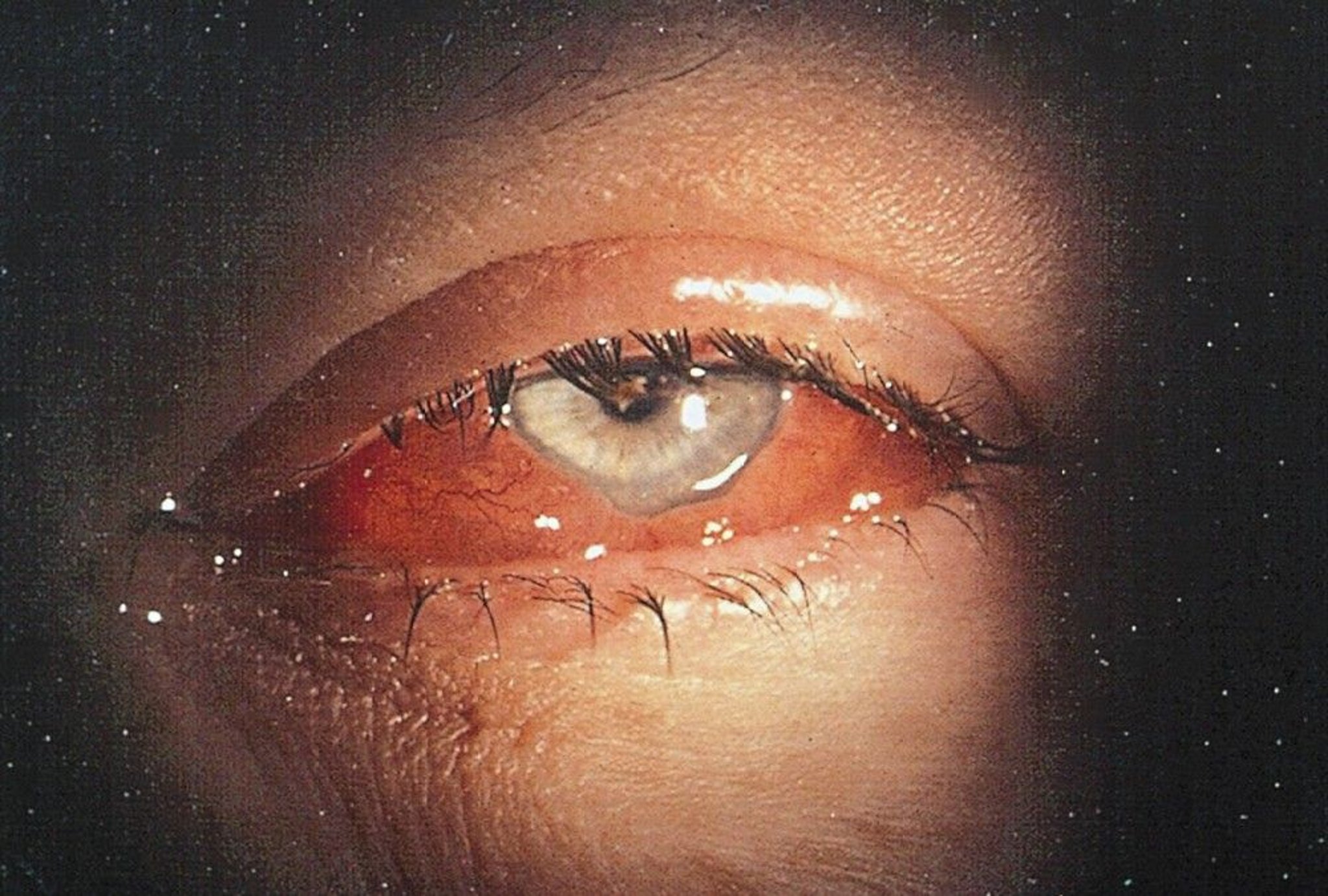
Edema of the bulbar conjunctiva results in a diffusely translucent, bluish, thickened conjunctiva. Gross edema with ballooning of the conjunctiva, often leading to prolapse of conjunctiva, is known as chemosis.

© Springer Science+Business Media
Edema of the tarsal conjunctiva (typical of allergic conjunctivitis) results in fine, minute projections (papillae), giving the tarsal conjunctiva a velvety appearance.
Hyperplasia of lymphoid follicles in the conjunctiva can occur in viral conjunctivitis or chlamydial conjunctivitis. It appears as small bumps with pale centers, resembling cobblestones. It occurs most commonly in the inferior tarsal conjunctiva.


