


Etiology
Major risk factors include
Age
Other risk factors include
Occlusion may also be idiopathic. The condition is uncommon among young people. Occlusion may affect a branch of the retinal vein or the central retinal vein.
Neovascularization (abnormal new vessel formation) of the retina or iris (rubeosis iridis) occurs in about 16% of patients with central retinal vein occlusion and can result in secondary (neovascular) glaucoma, which can occur weeks to months after occlusion. Vitreous hemorrhage may result from retinal neovascularization.
Symptoms and Signs
Painless vision loss is usually sudden but it can also occur gradually over a period of days to weeks. Funduscopy reveals hemorrhages throughout the retina, engorged (dilated) and tortuous retinal veins, and, usually, significant retinal edema. These changes are typically diffuse if obstruction involves the central retinal vein and are limited to one quadrant if obstruction involves only a branch of the central retinal vein.

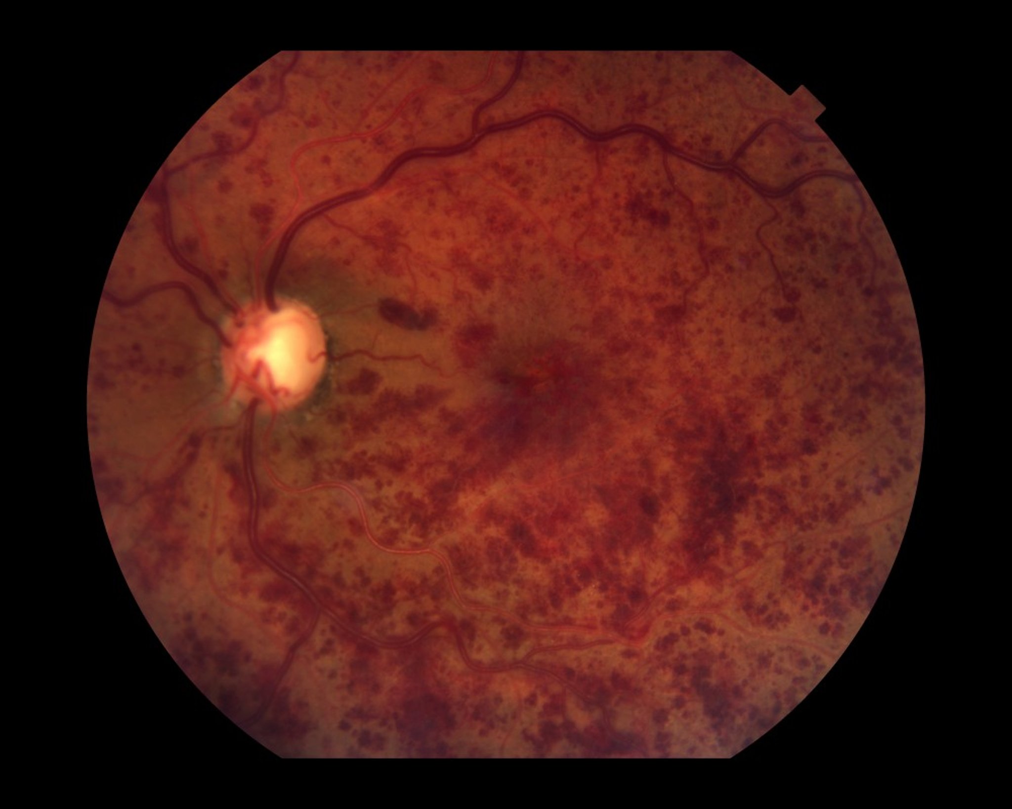
Paul Whitten/SCIENCE PHOTO LIBRARY
Diagnosis
Funduscopy
Color fundus photography
Optical coherence tomography
hypertension and glaucoma and tested for diabetes. Young patients are tested for increased blood viscosity (with a complete blood count and clotting factors as deemed necessary).
Prognosis
Most patients have some visual deficit. In mild cases, there can be spontaneous improvement to near-normal vision over a variable period of time. Visual acuity at presentation is a good indicator of final vision. If visual acuity is at least 20/40, visual acuity will likely remain good, occasionally near normal. If visual acuity is worse than 20/200, it will remain at that level or worsen in 80% of patients. Central retinal vein occlusions rarely recur.
Treatment
For some cases of macular edema with branch retinal vein occlusion, focal laser photocoagulation
Panretinal laser photocoagulation if neovascularization develops
If retinal or anterior segment neovascularization develops secondary to central or branch retinal vein occlusion, panretinal laser photocoagulation should be done promptly to decrease vitreous hemorrhage and prevent neovascular glaucoma.
Key Points
Retinal vein occlusion involves blockage by a thrombus.
Patients have painless loss of vision that is typically sudden and may have risk factors (eg, older age, hypertension).
Focal laser photocoagulation is useful in some cases of macular edema secondary to a branch retinal vein occlusion, and panretinal laser photocoagulation should be done for retinal or anterior segment neovascularization.


