


Excess tearing may cause a sensation of watery eyes or result in tears falling down the cheek (epiphora).
Pathophysiology of Tearing
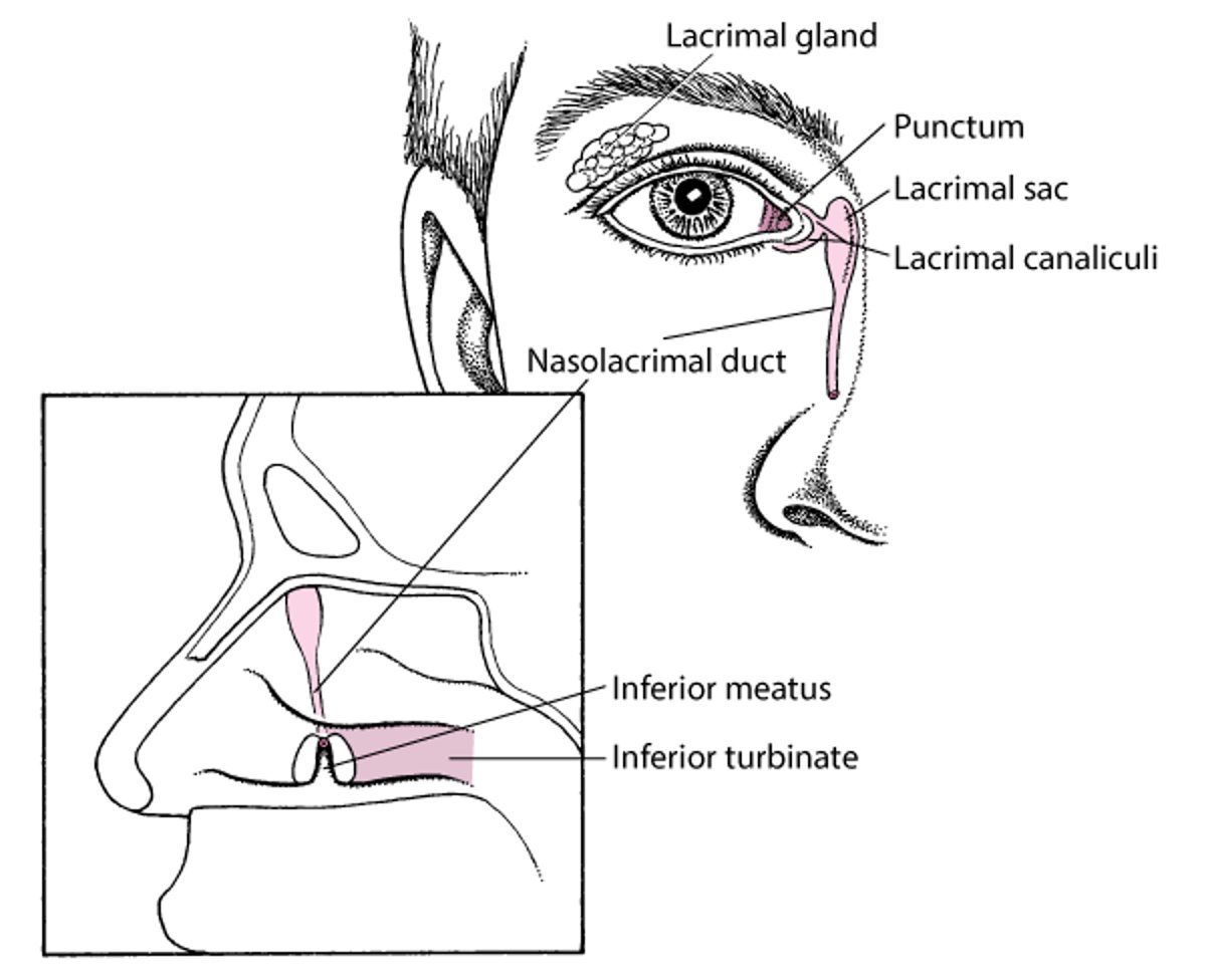
Tears are produced in the lacrimal gland and drain through the upper and lower puncta into the canaliculi and then into the lacrimal sac and nasolacrimal duct (see figure Anatomy of the Lacrimal System). Obstruction of tear drainage can lead to stasis and infection. Recurrent infection of the lacrimal sac (dacryocystitis) can sometimes spread, potentially leading to orbital cellulitis.
Anatomy of the Nasolacrimal System

Etiology of Tearing
Overall, the most common causes of tearing are
Upper respiratory infection
Allergic rhinitis
Tearing can be caused by increased tear production or decreased nasolacrimal drainage. In many patients, the cause of tearing can be multifactorial.
Increased tear production
The most common causes are
Upper respiratory infection
Dry eyes (reflex tearing produced in response to dryness of the ocular surface)
Any disorder causing conjunctival or corneal irritation can increase tear production (see table Some Causes of Tearing). However, most patients with corneal disorders that cause excess tearing (eg, corneal abrasion, corneal ulcer, corneal foreign body, keratitis) or with primary angle-closure glaucoma or anterior uveitis present with eye symptoms other than tearing (eg, eye pain, redness). Most people who have been crying do not present for evaluation of tearing.
Decreased nasolacrimal drainage
The most common causes are
Idiopathic age-related nasolacrimal duct stenosis
Nasolacrimal drainage system obstruction may be caused by strictures, tumors, or foreign bodies (eg, stones, often associated with subclinical infection by Actinomyces). Obstruction can also be a congenital malformation. Many disorders and medications can cause stricture or obstruction of nasolacrimal drainage.
Other causes of nasolacrimal drainage stricture or obstruction include
Burns
Chemotherapy drugs
Infection, including canaliculitis (eg, caused by Staphylococcus aureus, Actinomyces, Streptococcus, Pseudomonas, herpes zoster virus, herpes simplex conjunctivitis, infectious mononucleosis, human papillomavirus, Ascaris, leprosy, tuberculosis)
Inflammatory disorders (sarcoidosis, granulomatosis with polyangiitis)
Injuries (eg, nasoethmoid fractures; nasal, orbital, or endoscopic sinus surgery)
Obstruction of nasal outlet despite an intact nasolacrimal system (eg, upper respiratory infection, allergic rhinitis, sinusitis)
Radiation therapy
Evaluation of Tearing
History
History of present illness addresses the duration, onset, and severity of symptoms, including whether tears drip down the cheek (true epiphora). The effects of weather, environmental humidity, and cigarette smoke are ascertained.
Review of symptoms should seek symptoms of possible causes, including itching, rhinorrhea, or sneezing, particularly when occurring perennially or after exposure to specific potential allergens (allergic reaction); eye irritation or pain (blepharitis, corneal abrasion, irritant chemicals); and pain near the medial canthus (dacryocystitis). Other symptoms are of lower yield but should be sought; they include positional headache, purulent rhinorrhea, nocturnal cough, and fever (sinusitis, granulomatosis with polyangiitis); rash (Stevens-Johnson syndrome); cough, dyspnea, and chest pain (sarcoidosis); and epistaxis, hemoptysis, polyarthralgias, and myalgias (granulomatosis with polyangiitis).
Past medical history asks about known disorders that can cause tearing, including granulomatosis with polyangiitis, sarcoidosis, and cancer treated with chemotherapy medications; disorders that cause dry eyes (eg, rheumatoid arthritis, sarcoidosis, Sjögren syndrome
Physical examination
Examination focuses on the eye and surrounding structures.
The nose is examined for congestion, purulence, and bleeding.
Red flags
The following findings are of particular concern:
Repeated, unexplained episodes of tearing
Hard mass in or near the nasolacrimal drainage structures
Interpretation of findings
Findings that suggest obstruction of nasolacrimal drainage include:
Tears running down the cheek (true epiphora)
Absence of a different specific cause
For a list of common causes that may be evident from the Ophthalmic evaluation, see table Some Causes of Tearing.
Testing
Testing is often unnecessary because the cause is usually evident from the examination.
Schirmer test with a large amount of wetting (eg, > 25 mm) suggests an evaporative dry eye as the etiology of tearing. Schirmer test with very little wetting (< 5.5 mm) suggests an aqueous tear-deficient dry eye. Usually, Schirmer test is done by an ophthalmologist to ensure it is done and interpreted correctly.
Imaging tests and procedures (dacryocystography, CT, nasal endoscopy) are sometimes useful to delineate abnormal anatomy when surgery is being considered or occasionally to detect an abscess. Recurrent infectious dacryocystitis can progress to more serious disorders such as orbital cellulitis.
Treatment of Tearing
Underlying disorders (eg, allergies, foreign bodies, conjunctivitis) are treated.
Congenital nasolacrimal duct obstruction often resolves spontaneously. In patients < 1 year, manual compression of the lacrimal sac 4 or 5 times a day may relieve the distal obstruction. After 1 year, the nasolacrimal duct may need probing with the patient under general anesthesia. If obstruction is recurrent, a temporary drainage tube may be inserted.
In acquired nasolacrimal duct obstruction, irrigation of the nasolacrimal duct may be therapeutic when underlying disorders do not respond to treatment. As a last resort, a passage between the lacrimal sac and the nasal cavity can be created surgically (dacryocystorhinostomy).
In cases of punctal or canalicular stenosis, dilation is usually curative. If canalicular stenosis is severe and bothersome, a surgical procedure that places a glass tube leading from the caruncle into the nasal cavity can be considered.
Geriatrics Essentials: Tearing
Idiopathic age-related nasolacrimal duct stenosis is the most common cause of unexplained epiphora in older patients; however, tumors should also be considered.
Key Points
If tears do not run down the cheek, dry eyes is often the cause.
If tears run down the cheek, obstruction of nasolacrimal drainage is likely.
Testing (dacryocystography, CT, nasal endoscopy) is often unnecessary but may be needed when surgery is being considered or occasionally to detect an abscess.


