


Lupus nephritis is glomerulonephritis caused by systemic lupus erythematosus (SLE). Clinical findings include hematuria, nephrotic-range proteinuria, and, in advanced stages, azotemia. Diagnosis is based on renal biopsy. Treatment is of the underlying disorder and usually involves corticosteroids and other immunosuppressant medications.
Lupus nephritis is diagnosed in about 50% of patients with SLE and typically develops within 1 year of diagnosis.
Pathophysiology of Lupus Nephritis
Pathophysiology involves immune complex deposition with development of glomerulonephritis. The immune complexes consist of
Nuclear antigens (especially DNA)
High-affinity complement-fixing IgG antinuclear antibodies
Antibodies to DNA
Subendothelial, intramembranous, subepithelial, or mesangial deposits are characteristic. Wherever immune complexes are deposited, immunofluorescence staining is positive for complement and for IgG, IgA, and IgM in varying proportions. Epithelial cells may proliferate, forming crescents.
Classification of lupus nephritis is based on histologic findings (see table Classification of Lupus Nephritis).
Antiphospholipid syndrome nephropathy
This syndrome may occur with or without lupus nephritis in up to one third of patients with SLE. The syndrome occurs in the absence of any other autoimmune process in 30 to 50% of affected patients. In antiphospholipid antibody syndrome, circulating lupus anticoagulant causes microthrombi, endothelial damage, and cortical ischemic atrophy. Antiphospholipid syndrome nephropathy increases a patient’s risk of hypertension and renal insufficiency or failure compared with lupus nephritis alone.
Symptoms and Signs of Lupus Nephritis
The most prominent symptoms and signs are those of SLE; patients who present with renal disease may have edema, hypertension, or a combination.
Diagnosis of Lupus Nephritis
Urinalysis and serum creatinine (all patients with SLE)
Diagnosis is suspected in all patients with SLE, particularly in patients who have proteinuria, microscopic hematuria, red blood cell (RBC) casts, or hypertension. Diagnosis is also suspected in patients with unexplained hypertension, elevated serum creatinine levels, or abnormalities on urinalysis who have clinical features suggesting SLE.
Urinalysis is done and serum creatinine is measured. Elevated anti–double-stranded-DNA (anti-dsDNA) antibody titers and low complement (C3 and C4) levels often indicate active lupus nephritis and support the diagnosis.
If the aforementioned studies are abnormal, renal biopsy is usually done to confirm the diagnosis and classify the disorder histologically. Histologic classification helps determine prognosis and direct treatment.
Some of the histologic subtypes are similar to other glomerulopathies; eg, membranous lupus nephritis is histologically similar to idiopathic membranous nephropathy and diffuse proliferative lupus nephritis is histologically similar to type I membranoproliferative glomerulonephritis. Overlap between these categories is substantial, and patients may progress from one class to another.

Image provided by Agnes Fogo, MD, and the American Journal of Kidney Diseases' Atlas of Renal Pathology (see www.ajkd.org).
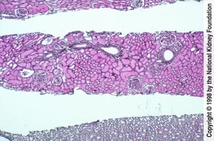
Image provided by Agnes Fogo, MD, and the American Journal of Kidney Diseases' Atlas of Renal Pathology (see www.ajkd.org).
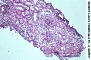
Image provided by Agnes Fogo, MD, and the American Journal of Kidney Diseases' Atlas of Renal Pathology (see www.ajkd.org).
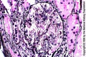
Image provided by Agnes Fogo, MD, and the American Journal of Kidney Diseases' Atlas of Renal Pathology (see www.ajkd.org).

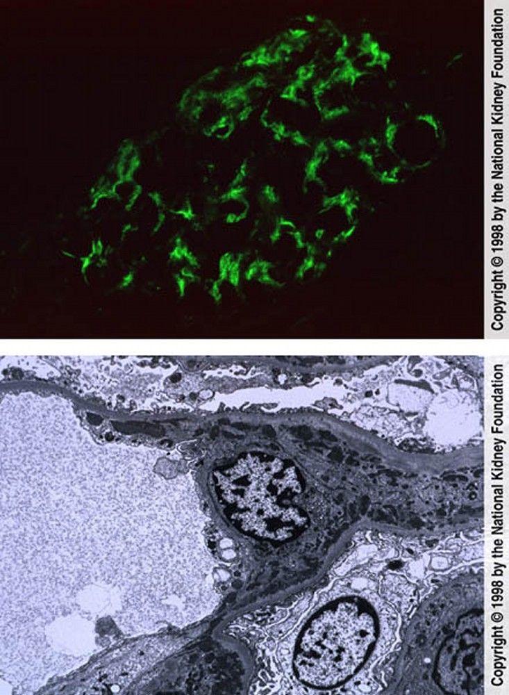
Image provided by Agnes Fogo, MD, and the American Journal of Kidney Diseases' Atlas of Renal Pathology (see www.ajkd.org).

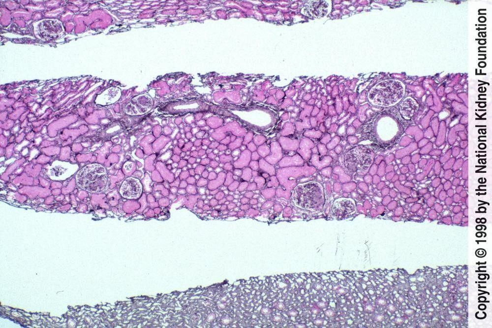
Image provided by Agnes Fogo, MD, and the American Journal of Kidney Diseases' Atlas of Renal Pathology (see www.ajkd.org).


Image provided by Agnes Fogo, MD, and the American Journal of Kidney Diseases' Atlas of Renal Pathology (see www.ajkd.org).
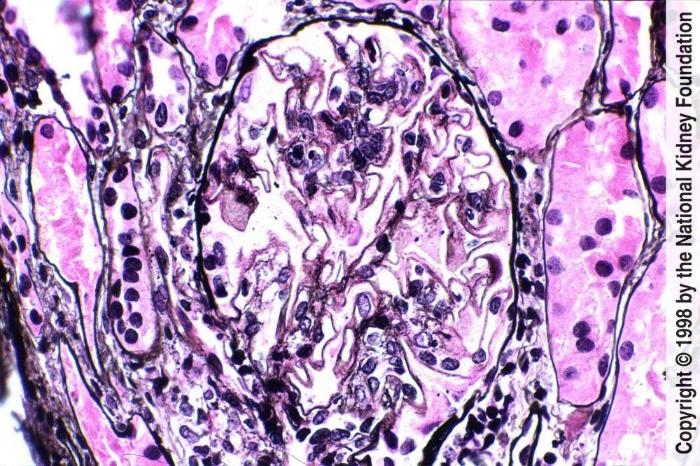
Image provided by Agnes Fogo, MD, and the American Journal of Kidney Diseases' Atlas of Renal Pathology (see www.ajkd.org).
Renal function and SLE activity should be monitored regularly. A rising serum creatinine level reflects deteriorating renal function, while falling serum C3 and C4 levels or a rising anti-dsDNA antibody titer suggests increased disease activity.
Treatment of Lupus Nephritis
Angiotensin inhibition for hypertension or proteinuria
Kidney transplantation for patients with end-stage kidney disease
Angiotensin inhibitionhypertension (eg, blood pressure [BP] > 130/80 mm Hg) or proteinuria. Also, dyslipidemia and risk factors for atherosclerosis should be treated aggressively.
Immunosuppression
Treatment is guided by the histologic classification of the lupus nephritis, the degree of disease activity and chronicity, and the presence of concomitant kidney disorders.
Activity is estimated by the activity score as well as clinical criteria (eg, urine sediment, increasing urine protein, increasing serum creatinine). Many experts believe that a mild to moderate chronicity score, because it suggests reversibility, should provoke more aggressive therapy than a more severe chronicity score. Nephritis with the potential for deterioration and for reversibility is usually class III or IV; it is unclear whether class V nephritis warrants aggressive treatment.
The activity score describes the degree of inflammation. The score is based on cellular proliferation, fibrinoid necrosis, cellular crescents, hyaline thrombi, wire loop lesions, glomerular leukocyte infiltration, and interstitial mononuclear cell infiltration. The activity score is less well correlated with disease progression and is used, rather, to help identify active nephritis.
The chronicity index describes the degree of scarring. It is based on presence of glomerular sclerosis, fibrous crescents, tubular atrophy, and interstitial fibrosis. The chronicity index predicts progression of lupus nephritis to renal failure. A mild to moderate chronicity score suggests at least partially reversible disease, whereas more severe chronicity scores may indicate irreversible disease.
Treatment for proliferative lupus nephritis combines corticosteroids with other immunosuppressants (1, 2).
Induction therapy for focal or diffuse lupus nephritis34, 5
maintenancetherapy
For patients with pure lupus membranous nephropathy, there is a lack of consensus regarding the role of immunosuppressive therapy, and some experts limit its use depending on the degree of proteinuria and findings on kidney biopsy. However, patients who have concurrent lupus membranous nephropathy and focal or diffuse lupus nephritis are treated using the same approach as for focal or diffuse lupus nephritis alone.
Other treatments
Anticoagulation is of theoretical benefit for patients with antiphospholipid syndrome nephropathy, but the value of such treatment has not been established. However, patients with antiphospholipid syndrome (APS) and a definite thrombotic event should be anticoagulated in accordance with preferred agents for APS.
Kidney transplantation is an option for patients with end-stage kidney disease due to lupus nephritis. Recurrent disease in the graft is uncommon (< 5%), but risk may be increased in Black people, females, and younger patients.
Treatment references
1. Fanouriakis A, Kostopoulou M, Cheema K, et al: 2019 Update of the Joint European League Against Rheumatism and European Renal Association-European Dialysis and Transplant Association (EULAR/ERA-EDTA) recommendations for the management of lupus nephritis. Ann Rheum Dis 9(6):713-723, 2020. doi: 10.1136/annrheumdis-2020-216924
2. Rovin BH, Caster DJ, Cattran DC, et al: Management and treatment of glomerular diseases (part 2): conclusions from a Kidney Disease: Improving Global Outcomes (KDIGO) Controversies Conference. Kidney Int 95(2):281-295, 2019. doi: 10.1016/j.kint.2018.11.008
3. Tunnicliffe DJ, Palmer SC, Henderson L, et al: Cochrane Database Syst Rev ;6(6):CD002922, 2018. doi: 10.1002/14651858.CD002922.pub4
4. Rovin BH, Onno Teng YK, Ginzler EM, et alLancet 397(10289):2070-2080, 2021. doi: 10.1016/S0140-6736(21)00578-X
5. Rovin BH, Furie R, Onno Teng YK, et alKidney Int 101(2):403-413, 2022. doi: 10.1016/j.kint.2021.08.027
Prognosis for Lupus Nephritis
Class of nephritis influences renal prognosis (see table Classification of Lupus Nephritis), as do other renal histologic features. Kidney biopsies are scored with a chronicity index and with a semiquantitative activity score.
Black patients with lupus nephritis are also at higher risk of progression to end-stage kidney disease.
Patients with lupus nephritis are at high risk of cancers, primarily B-cell lymphomas. Risk of atherosclerotic complications (eg, coronary artery disease, ischemic stroke) is also high because of frequent vasculitis, hypertension, dyslipidemia, and use of corticosteroids.
Key Points
Nephritis, although clinically evident in only 50%, probably occurs in > 90% of patients with SLE.
Obtain urinalysis and measure serum creatinine in all patients with SLE and renal biopsy if an unexplained abnormality is found in either, particularly in the presence of low C3 and C4 and elevated dsDNA.
Initiate angiotensin inhibition for even mild hypertension and treat atherosclerotic risk factors aggressively.


