|
Tubulointerstitial nephritis is primary injury to renal tubules and interstitium resulting in decreased renal function. The acute form is most often due to allergic drug reactions or to infections. The chronic form occurs with a diverse array of causes, including genetic or metabolic disorders, obstructive uropathy, and chronic exposure to environmental toxins or to certain drugs and herbs. Diagnosis is suggested by history and urinalysis and often confirmed by biopsy. Treatment and prognosis vary by the etiology and potential reversibility of the disorder at the time of diagnosis.
Etiology
Tubulointerstitial nephritis can be primary, but a similar process can result from glomerular damage (see Glomerular Disorders: Overview of Glomerular Disorders) or renovascular disorders (see Renovascular Disorders).
Primary tubulointerstitial nephritis may be
Acute tubulointerstitial nephritis (ATIN):
ATIN involves an inflammatory infiltrate and edema affecting the renal interstitium that often develops over days to months. Over 95% of cases result from infection or an allergic drug reaction. A syndrome of ATIN associated with uveitis (renal-ocular syndrome) also occurs and is idiopathic. ATIN causes acute renal insufficiency or failure; severe cases, delayed therapy, or continuance of an offending drug can lead to permanent injury with chronic renal failure.
Chronic tubulointerstitial nephritis (CTIN):
CTIN arises when chronic tubular insults cause gradual interstitial infiltration and fibrosis, tubular atrophy and dysfunction, and a gradual deterioration of renal function, usually over years. Glomerular involvement (glomerulosclerosis) is much more common in CTIN than in ATIN. Causes of CTIN are myriad; they include immunologically mediated disorders, infections, reflux or obstructive nephropathy, drugs, and other disorders. CTIN due to toxins, metabolic derangements, hypertension, and inherited disorders results in symmetric and bilateral disease; with other causes, renal scarring may be unequal and involve only one kidney. Some well-characterized forms of CTIN include analgesic, metabolic, heavy metal, and reflux nephropathy and myeloma kidney (hereditary cystic kidney diseases are discussed in see Cystic Kidney Disease: Overview of Cystic Kidney Disease).
|
Table 2
|
 |  |  |
| Causes of Acute Tubulointerstitial Nephritis |
|
Cause
|
Examples
|
|
Drugs*
|
|
Antibiotics
|
β-Lactam antibiotics (the most common cause, particularly methicillin†)
Ciprofloxacin
Ethambutol
Indinavir
Isoniazid
Macrolides
Minocycline
Rifampin
Tetracycline
Trimethoprim/ sulfamethoxazole
Vancomycin
|
|
Anticonvulsants
|
Carbamazepine
Phenobarbital
Phenytoin
Valproate
|
|
Diuretics
|
Bumetanide
Furosemide
Thiazides
Triamterene
|
|
NSAIDs
|
Diclofenac
Ibuprofen
Indomethacin
Naproxen
|
|
Other
|
Allopurinol
Aristocholic acid‡
Captopril
Cimetidine
Interferon alfa
Lansoprazole
Mesalazine
Omeprazole
Ranitidine
|
|
Metabolic
|
|
Hyperoxalaturia
|
Ethylene glycol poisoning
|
|
Hyperuricosuria
|
Tumor lysis syndrome
|
|
Renal parenchymal infection
|
|
Bacterial
|
Brucella sp
Corynebacterium diphtheriae
Legionella sp
Leptospira sp
Mycobacterium sp
Mycoplasma sp
Rickettsia sp
Salmonella sp
Staphylococci
Streptococci
Treponema pallidum
Yersinia sp
|
|
Fungal
|
Candida sp
|
|
Parasitic
|
Toxoplasma gondii
|
|
Viral
|
Cytomegalovirus
Epstein-Barr virus
Hantavirus
Hepatitis C virus
HIV
Mumps
Polyomavirus
|
|
Other
|
|
Idiopathic without and with uveitis
|
—
|
|
Immunologic
|
Cryoglobulinemia
IgA nephropathy
Renal transplant rejection
Sarcoidosis
Sjögren's syndrome
SLE (rare)
Wegener's granulomatosis
|
|
Neoplastic
|
Lymphoma
Myeloma
|
|
*Most common causative drugs are listed; > 120 drugs are implicated.
|
|
† No longer available in the US.
|
|
‡Contained in some medicinal herbs used in traditional Chinese medicine.
|
|
|
Table 3
|
 |  |  |
| Causes of Chronic Tubulointerstitial Nephritis |
|
Cause
|
Examples
|
|
Balkan nephropathy
|
—
|
|
Cystic diseases
|
Acquired cystic disease
Medullary cystic disease
Medullary sponge kidney
Nephronophthisis
Polycystic kidney disease*
|
|
Drugs
|
Analgesics*
Antineoplastics (cisplatin and nitrosourea)
Immunosuppressants (cyclosporine* and tacrolimus)
Lithium*
|
|
Granulomatous
|
Inflammatory bowel disease
Sarcoidosis
TB
Wegener's granulomatosis
|
|
Hematologic
|
Aplastic anemia
Leukemia
Lymphoma
Multiple myeloma*
Sickle cell anemia
|
|
Hereditary nephropathy associated with hyperuricemia and gout
|
—
|
|
Idiopathic
|
—
|
|
Immunologic
|
Amyloidosis
Cryoglobulinemia
Goodpasture's syndrome
IgA nephropathy
Renal transplant rejection
Sarcoidosis
Sjögren's syndrome
SLE
|
|
Infection
|
Renal parenchymal: Pyelonephritis, Hantavirus—Puumula type infection (nephropathia epidemica)
Systemic
|
|
Mechanical
|
Obstructive uropathy
Reflux nephropathy*
|
|
Metabolic
|
Chronic hypokalemia
Cystinosis
Fabry's disease
Hypercalcemia, hypercalciuria
Hyperoxaluria
Hyperuricemia*, hyperuricosuria
|
|
Radiation nephritis
|
—
|
|
Toxins
|
Aristocholic acid†
Heavy metals (eg, arsenic, bismuth, cadmium, chromium, copper, gold, iron, lead, mercury, uranium)
|
|
Vascular
|
Atheroembolism
Hypertension
Renal vein thrombosis
|
|
*Common causes.
|
|
†Contained in some medicinal herbs used in traditional Chinese medicine.
|
|
Symptoms and Signs
ATIN:
Symptoms and signs of ATIN may be nonspecific and are often absent unless symptoms and signs of renal failure develop. Many patients develop polyuria and nocturia (due to a defect in urinary concentration and Na reabsorption). Symptom onset may be as long as several weeks after initial toxic exposure or as soon as 3 to 5 days after a 2nd exposure; extremes in latency range from 1 day with rifampin to 18 mo with an NSAID. Fever and urticarial rash are characteristic early manifestations of drug-induced ATIN, but the classically described triad of fever, rash, and eosinophilia is insensitive. Abdominal pain, weight loss, and bilateral renal masses (caused by interstitial edema) may also occur in ATIN and with fever may mistakenly suggest renal cancer or polycystic kidney disease. Peripheral edema and hypertension are uncommon unless renal insufficiency or renal failure occurs.
CTIN:
Symptoms and signs are generally absent in CTIN unless renal failure develops. Edema usually is not present, and BP is normal or only mildly elevated in the early stages. Polyuria and nocturia may develop.
Diagnosis
Few clinical and routine laboratory findings are specific. Thus, suspicion should be high when the following are present:
Other tests (eg, imaging) may be necessary to differentiate ATIN or CTIN from other disorders. Renal biopsy is sometimes done.
ATIN:
Signs of active kidney inflammation (active urinary sediment), including RBCs, WBCs, and WBC casts, and absence of bacteria on culture (sterile pyuria) are typical; marked hematuria and dysmorphic RBCs are uncommon. Eosinophiluria has a positive predictive value of 50% (specificity of about 85 to 93%) and a negative predictive value of up to 90% for ATIN (sensitivity of about 63 to 91%). Thus, the presence of urinary eosinophils is not diagnostic, but their absence significantly decreases the likelihood of the diagnosis. Proteinuria is usually minimal but may reach nephrotic range with combined ATIN-glomerular disease induced by NSAIDs, ampicillin, rifampin, interferon alfa, or ranitidine. Blood test findings of tubular dysfunction include hypokalemia (caused by a defect in K reabsorption) and a nonanion gap metabolic acidosis (caused by a defect in HCO3 reabsorption or acid excretion).
Ultrasonography, radionuclide scanning, or both may be needed to differentiate ATIN from other causes of acute renal failure, such as acute tubular necrosis. In ATIN, ultrasonography may show kidneys that are greatly enlarged and echogenic because of interstitial inflammatory cells and edema. Radionuclide scans may show kidneys avidly taking up radioactive gallium-67 or radionuclide-labeled WBCs. Positive scans strongly suggest ATIN (and indicate that acute tubular necrosis is less likely), but a negative scan does not exclude ATIN.
Renal biopsy is usually reserved for patients with the following:
In ATIN, glomeruli are usually normal. The earliest finding is interstitial edema, typically followed by interstitial infiltration with lymphocytes, plasma cells, eosinophils, and a few PMNs. In severe cases, inflammatory cells can be seen invading the space between the cells lining the tubular basement membrane (tubulitis); in other specimens, granulomatous reactions resulting from exposure to methicillin, sulfonamides, mycobacteria, or fungi may be seen. The presence of noncaseating granulomas suggests sarcoidosis. Immunofluorescence or electron microscopy seldom reveals any pathognomonic changes.
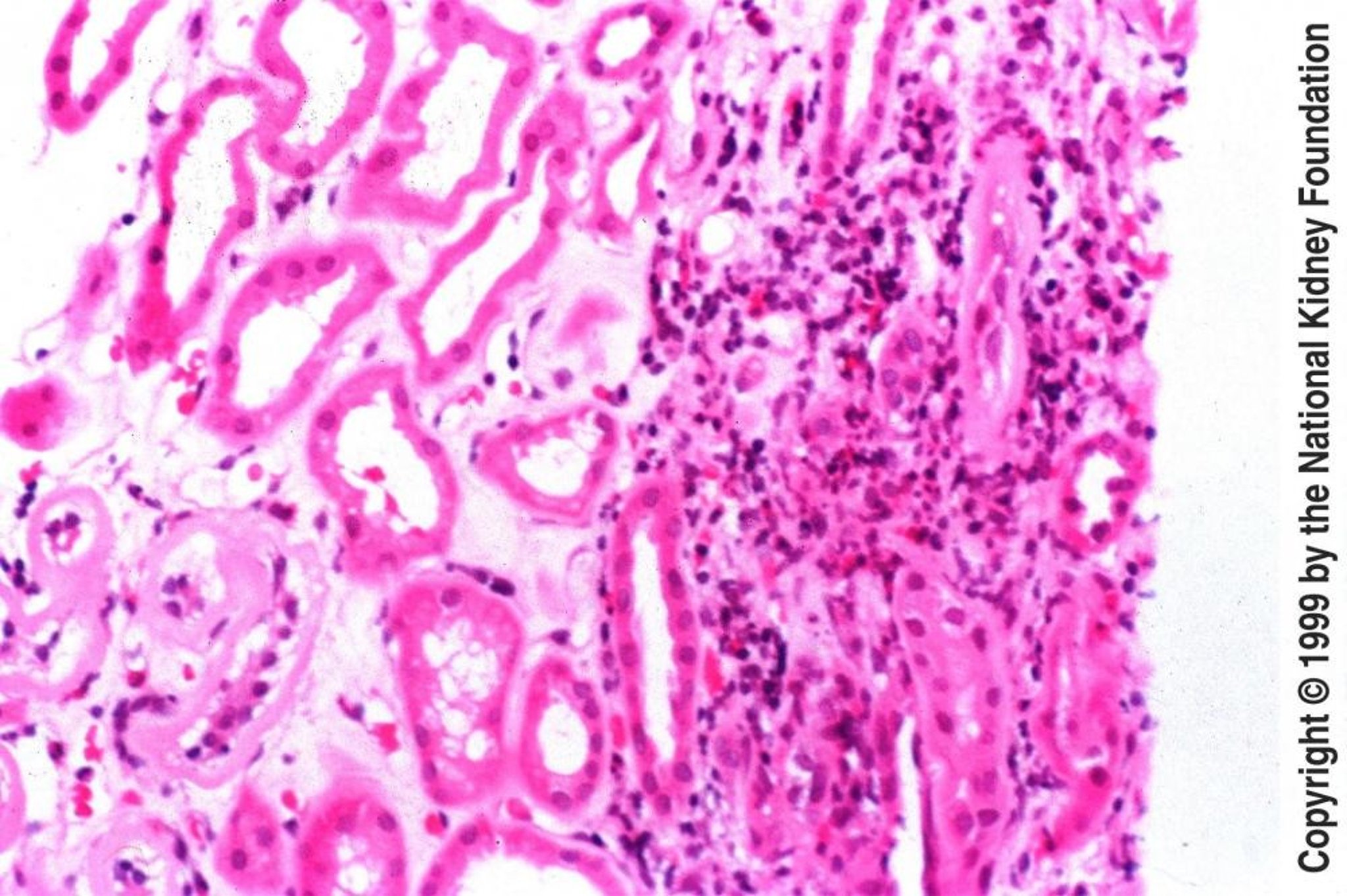
CTIN:
Findings of CTIN are generally similar to those of ATIN, although urinary RBCs and WBCs are uncommon. Because CTIN is insidious in onset and interstitial fibrosis is common, imaging tests may show small kidneys with evidence of scarring and asymmetry.
In CTIN, renal biopsy is not often done for diagnostic purposes but has helped characterize the nature and progression of tubulointerstitial disease. Glomeruli vary from normal to completely destroyed. Tubules may be absent or atrophied. Tubular lumina vary in diameter but may show marked dilation, with homogeneous casts. The interstitium contains varying degrees of inflammatory cells and fibrosis. Nonscarred areas appear almost normal. Grossly, the kidneys are small and atrophic.
Prognosis
In drug-induced ATIN, renal function usually recovers within 6 to 8 wk when the offending drug is stopped, although some residual scarring is common. Recovery may be incomplete, with persistent azotemia above baseline. When other factors cause ATIN, histologic changes usually are reversible if the cause is recognized and removed; however, some severe cases progress to fibrosis and renal failure. Regardless of cause, irreversible injury is suggested by the following:
In CTIN, prognosis depends on the cause and on the ability to recognize and stop the process before irreversible fibrosis occurs. Many genetic (eg, cystic kidney disease), metabolic (eg, cystinosis), and toxic (eg, heavy metal) causes may not be modifiable, in which case CTIN usually evolves to end-stage renal disease.
Treatment
Treatment of both ATIN and CTIN is management of the cause. For immunologically induced disease in ATIN and perhaps CTIN and sometimes drug-induced ATIN, corticosteroids (eg, prednisone 1 mg/kg po once/day with gradual tapering of the dose over 4 to 6 wk) may accelerate recovery. Treatment of CTIN often requires supportive measures such as controlling BP and treating anemia associated with kidney disease. In patients with CTIN and progressive renal injury, ACE inhibitors or angiotensin II receptor blockers may slow disease progression.
Analgesic Nephropathy
Analgesic nephropathy (AN) is CTIN caused by cumulative lifetime use of large amounts (eg, ≥ 2 kg) of certain analgesics.
AN was originally described in conjunction with overuse of combination analgesics containing phenacetin (typically with aspirin, acetaminophen, codeine, or caffeine). However, despite removal of phenacetin from the market, AN continued to occur. Studies to identify the causal agent are equivocal, but acetaminophen, aspirin, and other NSAIDs have been implicated. Mechanism is unclear. Whether COX-2 inhibitors cause AN is not known, but these drugs probably can cause ATIN and nephrotic syndrome due to minimal change disease or membranous nephropathy.
AN predominates in women (peak incidence, 50 to 55 yr) and, in the US, is responsible for 3 to 5% of cases of end-stage renal disease (13 to 20% in Australia and South Africa).
Patients present with renal insufficiency and usually non-nephrotic proteinuria with a bland urinary sediment or sterile pyuria. Hypertension, anemia, and impaired urinary concentration are common once renal insufficiency develops. Flank pain and hematuria are signs of papillary necrosis that occur late in the course of disease. Chronic complaints of musculoskeletal pain, headache, malaise, and dyspepsia may be related to long-term analgesic use rather than AN.
Diagnosis is based on history of chronic analgesic use and noncontrast CT. CT signs of AN are the following:
The combination of these findings has a sensitivity of 85% and a specificity of 93% for early diagnosis, but these specificity and sensitivity numbers are based on studies done when use of phenacetin-containing analgesics was widespread.
Renal function stabilizes when analgesics are stopped unless renal insufficiency is advanced, in which case it may progress to renal failure. Patients with AN are at greater risk of transitional cell carcinomas of the urinary tract.
Metabolic Nephropathies
Tubulointerstitial disorders can result from several metabolic disturbances.
Acute urate nephropathy:
This disorder is not a true form of ATIN but rather an intraluminal obstructive uropathy caused by uric acid crystal deposition within the lumen of renal tubules; acute oliguric or anuric kidney injury results. Causes include the following:
Typically, no symptoms are present. Diagnosis is suspected when acute kidney injury occurs in patients with marked hyperuricemia (> 15 mg/dL). Urinalysis results may be normal or may show urate crystals.
Prognosis for complete recovery of renal function is excellent if treatment is initiated rapidly. Treatment is usually with allopurinol plus aggressive IV hydration in patients with normal cardiac and renal function. Supportive measures are indicated. Hemodialysis may be recommended to remove excess circulating urate in severe cases where diuresis cannot be induced with a loop diuretic and IV saline.
Prevention is indicated for patients at high risk (eg, those at risk of tumor lysis syndrome). Prevention is by use of allopurinol 300 mg po bid to tid plus saline loading to maintain a urine output > 2.5 L/day before chemotherapy or radiation therapy. Urate oxidase (rasburicase), which catalyzes urate to a much more soluble compound, is also preventive and is being more commonly used in patients with severe hyperuricemia. However, patients given rasburicase must be carefully monitored because the drug must be given IV and can cause anaphylaxis, hemolysis, and other adverse effects.
Chronic urate nephropathy:
This condition is CTIN caused by deposition of Na urate crystals in the medullary interstitium in patients with chronic hyperuricemia. Sequelae are chronic inflammation and fibrosis, with ensuing chronic renal insufficiency and renal failure. Chronic urate nephropathy was once common in patients with tophaceous gout but is now rare because gout is more often effectively treated. A bland urine sediment and hyperuricemia disproportionate to the degree of renal insufficiency (eg, urate > 9 mg/dL with serum creatinine < 1.5 mg/dL, or > 10 mg/dL with serum creatinine 1.5 to 2 mg/dL, and > 12 mg/dL with more advanced renal failure) are suggestive but nonspecific; many causes of tubulointerstitial diseases may have these findings, lead nephropathy being the most common. Treatment is that of hyperuricosuria (see Crystal-Induced Arthritides: Lowering the serum urate level).
Hyperoxaluria:
Hyperoxaluria is a common cause of nephrolithiasis but an uncommon cause of acute and chronic tubulointerstitial nephritis. Causes and prevention of hyperoxaluria are discussed elsewhere (see Urinary Calculi).
Hypercalcemia:
Hypercalcemia (see Electrolyte Disorders: Hypercalcemia) causes nephropathy by 2 mechanisms. Severe (> 12 mg/dL) temporary hypercalcemia may cause reversible renal insufficiency by renal vasoconstriction and natriuresis-induced volume depletion. Long-standing hypercalcemia and hypercalciuria lead to CTIN with calcification and necrosis of tubular cells, interstitial fibrosis, and calcification (nephrocalcinosis). Common associated findings include
Diagnosis is based on presence of hypercalcemia and unexplained renal insufficiency; nephrocalcinosis can be detected by ultrasonography or noncontrast CT. Treatment is management of hypercalcemia.
Chronic hypokalemia:
Chronic hypokalemia of a moderate to severe degree may cause nephropathy with impaired urinary concentration and vacuolation of proximal tubular cells and occasionally of distal tubular cells. Chronic interstitial inflammatory changes, fibrosis, and renal cysts have been found in renal biopsies of patients with hypokalemia of ≥ 1 mo. Treatment consists of correction of the underlying disorder and oral K supplements. Although the hypokalemia as well as the number and size of the cysts are reversible, the CTIN and renal insufficiency may be irreversible.
Heavy Metal Nephropathy
Exposure to heavy metals and other toxins can result in tubulointerstitial disorders.
Lead:
CTIN results as lead accumulates in proximal tubular cells. Short-term lead exposure causes proximal tubular dysfunction, including decreased urate secretion and hyperuricemia (urate is the substrate for saturnine gout), aminoaciduria, and renal glucosuria. Chronic lead exposure (ie, for 5 to ≥ 30 yr) causes progressive tubular atrophy and interstitial fibrosis, with renal insufficiency, hypertension, and gout. However, chronic low-level exposure may cause renal insufficiency and hypertension independent of tubulointerstitial disease. The following groups are at highest risk:
Exposed children may develop nephropathy during adulthood.
Hyperuricemia disproportionate to the degree of renal insufficiency (eg, urate > 9 mg/dL with serum creatinine < 1.5 mg/dL, or > 10 mg/dL with serum creatinine 1.5 to 2 mg/dL, and > 12 mg/dL with more advanced renal failure) and a bland urinary sediment are common. Diagnosis is usually made by measuring whole blood lead levels. Alternatively, x-ray fluorescence may be used to detect increased bone lead concentrations, which reflect high cumulative lead exposure. Treatment with chelation therapy (see Poisoning: Treatment) can stabilize renal function, but recovery may be incomplete.
Cadmium:
Cadmium from contaminated water, food, and tobacco and, mainly, from workplace exposures can cause nephropathy. It can also cause a glomerulopathy that is usually asymptomatic. Early manifestations of cadmium nephropathy are those of tubular dysfunction, including low molecular weight tubular proteinuria (eg, β2-microglobulin), aminoaciduria, and renal glucosuria. Symptoms and signs, when they occur, are attributable to chronic renal insufficiency and failure. Renal disease follows a dose-response curve. Diagnosis is likely with the following:
Treatment is elimination of cadmium exposure; chelation with Na calcium edetate (EDTA) may increase cadmium nephrotoxicity. Tubular proteinuria usually is irreversible.
Other heavy metals:
Other heavy metals that are nephrotoxic include
All cause tubular damage and dysfunction (eg, tubular proteinuria, aminoaciduria) as well as tubular necrosis, but glomerulopathies may predominate with some compounds (mercury, gold). Treatment involves removal of the patient from further exposure and either or both of the following:
Reflux Nephropathy
Reflux nephropathy is renal scarring presumably induced by vesicoureteral reflux of infected urine into the renal parenchyma. The diagnosis is suspected in children with UTI or a family history. Diagnosis is by voiding cystourethrography or radionuclide cystography. Children with moderate or severe reflux are treated with prophylactic antibiotics or surgical correction.
Traditionally, the mechanism of renal scarring has been thought to be chronic pyelonephritis. However, reflux is probably the single most important factor, and factors unrelated to reflux or pyelonephritis (eg, congenital factors) can contribute. Vesicoureteral reflux (VUR) affects about 1% of newborns and 30 to 45% of young children with a febrile UTI (see Miscellaneous Infections in Infants and Children: Urinary Tract Infection in Children (UTI)); it is common among children with renal scars and, for unknown reasons, is less common among black children than white children. Familial predisposition is common. Children with gross reflux (up to the renal pelvis plus ureteral dilatation) are at highest risk of scarring and subsequent renal failure.
Reflux requires incompetent ureterovesical valves or mechanical obstruction in the lower urinary tract. Young children with shorter intravesical portions of the ureter are most susceptible; normal growth usually results in spontaneous cessation of intrarenal and vesicoureteral reflux by age 5. New scars in children > 5 yr are unusual but may occur after acute pyelonephritis.
Symptoms and Signs
Few symptoms and signs other than occasional UTI are present in young children, and the diagnosis is often overlooked until adolescence, when patients present with polyuria, nocturia, hypertension, symptoms and signs of renal insufficiency, laboratory abnormalities, or a combination.
Diagnosis
The diagnosis may be suspected prenatally or postnatally. Diagnosis and staging of reflux nephropathy (prenatal or postnatal presentation) are made by a voiding cystourethrogram (VCUG), which can demonstrate the degree of ureteral dilatation. Radionuclide cystography (RNC) can also be used; it provides less anatomic detail than VCUG but involves less radiation exposure. Because these tests involve catheterization (and risk of UTI) as well as radiation exposure, thresholds for obtaining them can be controversial. Renal scarring is diagnosed with technetium-99m–labeled dimercaptosuccinic acid (DMSA) radionuclide scanning.
Prenatal presentation:
The diagnosis is suspected prenatally if ultrasonography, done because of a family history or for unrelated reasons, shows hydronephrosis; 10 to 40% of such patients are diagnosed postnatally with VUR. Some experts recommend VCUG or RNC only if family history is strong or if postnatal renal ultrasonography is markedly or persistently abnormal; however, it is not clear whether renal ultrasonography is sufficiently sensitive to detect VUR. DMSA scanning is typically done in neonates (as well as infants < 6 mo) who have worrisome UTIs (eg, febrile or recurrent UTIs).
Postnatal presentation:
VUR is suspected postnatally in patients with any of the following:
Laboratory abnormalities may include proteinuria, Na wasting, hyperkalemia, metabolic acidosis, renal insufficiency, or a combination. Testing for these patients is with RNC or VCUG. DMSA scanning may be done for infants or children with UTIs as listed above.
In older children in whom reflux is no longer active, a VCUG may not show reflux, although the DMSA scan shows scarring; cystoscopy can demonstrate evidence of previous reflux at ureteral orifices. Thus, DMSA scanning and cystoscopy may be done if prior reflux is suspected but not confirmed. Renal biopsy at this late stage shows CTIN and focal glomerulosclerosis, the cause of mild (1 to 1.5 g/day) to nephrotic-range proteinuria.
Treatment
Treatment is based on the unproven assumption that decreasing reflux and UTIs prevents renal scarring. Children with very mild VUR require no treatment, but they should be closely observed for symptoms of UTI. Children with moderate reflux are usually given antibiotics. However, drug therapy predisposes to new episodes of acute pyelonephritis, and it is not clear whether prophylactic antibiotics are more effective than close observation. Patients with severe reflux are at higher risk of renal insufficiency and are usually given antibiotic prophylaxis or undergo surgical interventions, including ureteral reimplantation or endoscopic injection of materials behind the ureter to prevent reflux (bladder contraction during voiding compresses the ureter between the bladder and the material). Incidence of new renal scars is similar in patients treated with surgery and with drugs.
Reflux spontaneously resolves in about 80% of young children within 5 yr.
Myeloma-Related Kidney Disease
Patients with multiple myeloma overproduce monoclonal Ig light chains (Bence Jones proteins); these light chains are filtered by glomeruli, are nephrotoxic, and can damage virtually all areas of the kidney parenchyma. Diagnosis is by urine tests (sulfosalicylic acid test or protein electrophoresis) or renal biopsy. Treatment focuses on the multiple myeloma and ensuring adequate urine flow.
Tubulointerstitial and glomerular damage are the most common types of renal damage. The mechanisms by which light chains damage nephrons directly are unknown. Hypercalcemia contributes to renal insufficiency by decreasing renal blood flow
Tubulointerstitial disease:
Light chains saturate the reabsorptive capacity of the proximal tubule, reach the distal nephron, and combine with filtered proteins and Tamm-Horsfall mucoprotein (secreted by cells of the thick ascending limb of Henle) to form obstructive casts. The term myeloma kidney or myeloma cast nephropathy generally refers to renal insufficiency caused by the tubulointerstitial damage that results. Factors that predispose to cast formation include the following:
Other types of tubulointerstitial lesions that occur with Bence Jones proteinuria include proximal tubular transport dysfunction causing Fanconi syndrome and light chain interstitial deposition with inflammatory infiltrates and active tubular damage.
Glomerulopathies:
Myeloma glomerulopathy has 2 common mechanisms: primary (AL) amyloidosis (see also Amyloidosis: Primary amyloidosis (AL)) and glomerular light chain or rarely heavy chain deposition. AL amyloidosis results in glomerular deposition of AL amyloid in the mesangial, subepithelial, or subendothelial areas or a combination. Amyloid deposition is with randomly oriented, nonbranching fibrils composed of the variable regions of λ light chains. Light chain deposition disease (LCDD), which also can occur with lymphoma and Waldenström's macroglobulinemia, is glomerular deposition of nonpolymerized light chains (ie, without fibrils), generally the constant regions of κ chains.
Rarely, a nonproliferative, noninflammatory glomerulopathy that causes nephrotic-range proteinuria can develop in advanced myeloma-related renal disease. A proliferative glomerulonephritis occasionally develops as an early form of LCDD with progression to membranoproliferative glomerulonephritis and nodular glomerulopathy reminiscent of diabetic nephropathy; nephrotic-range proteinuria is common.
Symptoms and Signs
Symptoms and signs are predominantly those of the myeloma (eg, skeletal pain, pathologic fractures, diffuse osteoporosis) and a normochromic-normocytic anemia.
Diagnosis
Diagnosis of myeloma-related kidney disease is suggested by the following combination of findings:
The diagnosis should be suspected even in patients without a history of or findings suggesting multiple myeloma. Diagnosis of light chain tubulointerstitial disease (myeloma kidney) is confirmed by a markedly positive urine sulfosalicylic acid test suggesting significant nonalbumin proteins, by urine protein electrophoresis (UPEP), or both. Diagnosis of glomerulopathy is confirmed by renal biopsy. Renal biopsy may demonstrate light chain deposition in 30 to 50% of patients with myeloma despite the absence of detectable serum or urine paraproteins by immunoelectrophoresis.
Prognosis
Kidney disease is a major predictor of overall prognosis in multiple myeloma. Prognosis is good for patients with tubulointerstitial and glomerular LCDD who receive treatment. Prognosis is worse for patients with AL amyloidosis, in whom amyloid deposition continues and progresses to renal failure in most cases. In either form without treatment, virtually all renal lesions progress to renal failure.
Treatment
Management of multiple myeloma (see Plasma Cell Disorders: Treatment), prevention of volume depletion, and maintenance of a high urine flow rate are the primary treatments. In addition, factors that worsen renal function (eg, hypercalcemia, hyperuricemia, use of nephrotoxic drugs) should be avoided or treated. Several measures are often recommended but are of unproved efficacy. Plasmapheresis may be tried to remove light chains. Alkalinization of the urine to help change the net charge of the light chain and reduce charge interaction with Tamm-Horsfall mucoprotein may make the light chains more soluble. Colchicine may be given to decrease secretion of Tamm-Horsfall mucoprotein into the lumen and to decrease the interaction with light chains, thus decreasing toxicity. Loop diuretics may be avoided to prevent volume depletion and high distal Na concentrations that can worsen myeloma-related kidney disease (loop diuretics are indicated if hypercalcemia is present).
Last full review/revision August 2009 by Navin Jaipaul, MD, MHS
Content last modified April 2010
|