


Sideroblastic anemias are a diverse group of anemias characterized by the presence of increased serum iron, ferritin, and transferrin saturation as well as ringed sideroblasts (erythroblasts with perinuclear iron-engorged mitochondria). Symptoms are those of anemia and include fatigue and lethargy. Diagnosis is with complete blood count, reticulocyte count, and peripheral blood smear as well as iron studies and bone marrow examination. Treatment requires stopping causative substances and giving vitamin supplements and erythropoietin.
(See also Overview of Decreased Erythropoiesis.)
Sideroblastic anemias may be
Acquired
Congenital
Acquired sideroblastic anemia is frequently associated with the myelodysplastic syndrome (but may be caused by medications or toxins) and causes a normocytic or macrocytic anemia.
Congenital sideroblastic anemia is caused by one of numerous X-linked or autosomal mutations and is usually a microcytic, hypochromic anemia but may be normocytic.
Sideroblastic anemias are iron-utilization anemias, which are characterized by inadequate mitochondrial utilization of iron due to impaired heme synthesis despite the presence of adequate or increased amounts of iron. Sideroblastic anemias are sometimes characterized by the presence of polychromatophilia (indicative of an increased number of reticulocytes) and stippled red blood cells (siderocytes) containing iron-laden granules (Pappenheimer bodies).
In both acquired and congenital sideroblastic anemia, heme synthesis is impaired due to the inability to incorporate iron into protoporphyrin IX, leading to the formation of ringed sideroblasts.
Acquired sideroblastic anemia
Most often, acquired sideroblastic anemias are part of a
Somatic mutations in genes involved in RNA splicing, most frequently SF3B1 (splicing factor 3b subunit 1), commonly occur. Acquired sideroblastic anemia occurs in adulthood.
Less common causes include
Deficiency of vitamin B6 (pyridoxine) or copper (possibly caused by zinc ingestion, which prevents absorption of copper in the gastrointestinal tract)
Toxins (including ethanol and lead)
Deficient reticulocyte production, intramedullary death of red blood cells (RBCs), and bone marrow erythroid hyperplasia (and dysplasia) occur. Although hypochromic RBCs are produced, other RBCs may be large, producing normocytic or macrocytic indices; if so, variation in RBC size (dimorphism) usually produces a high RBC distribution width (RDW).
Congenital sideroblastic anemia
Inherited forms of sideroblastic anemia are less common than acquired forms and usually occur in infancy or early childhood. The most common congenital sideroblastic anemia is an X-linked form caused by a germline mutation in ALAS2 (5'-aminolevulinate synthase 2), a gene involved in heme biosynthesis. Vitamin B6 (pyridoxine) is an essential cofactor for the enzyme produced by ALAS2
Numerous other X-linked, autosomal and mitochondrial forms have been identified with mutations in genes involved in heme synthesis or other mitochondrial enzymatic pathways (1).
RBCs are usually microcytic and hypochromic, but this is not always the case.
General reference
1. Ducamp S, Fleming MD: The molecular genetics of sideroblastic anemia. Blood 133:59–69, 2019. doi: 10.1182/blood-2018-08-815951
Diagnosis of Sideroblastic Anemias
Complete blood count (CBC), reticulocyte count, and peripheral blood smear
Iron studies (serum iron, serum ferritin, and transferrin saturation)
Bone marrow examination
Genetic testing for a suspected inherited or acquired mutation
Sideroblastic anemia is suspected in patients with microcytic anemia or a high RDW anemia, particularly with increased serum iron, serum ferritin, and transferrin saturation (see Iron Deficiency Anemia).
The peripheral smear shows RBC dimorphism. RBCs may appear stippled.
Bone marrow examination is necessary and reveals erythroid hyperplasia. Iron staining reveals the pathognomonic iron-engorged perinuclear mitochondria in developing RBCs (ringed sideroblasts). Other features of myelodysplastic syndrome, such as cytopenias and dysplasia, may be evident.

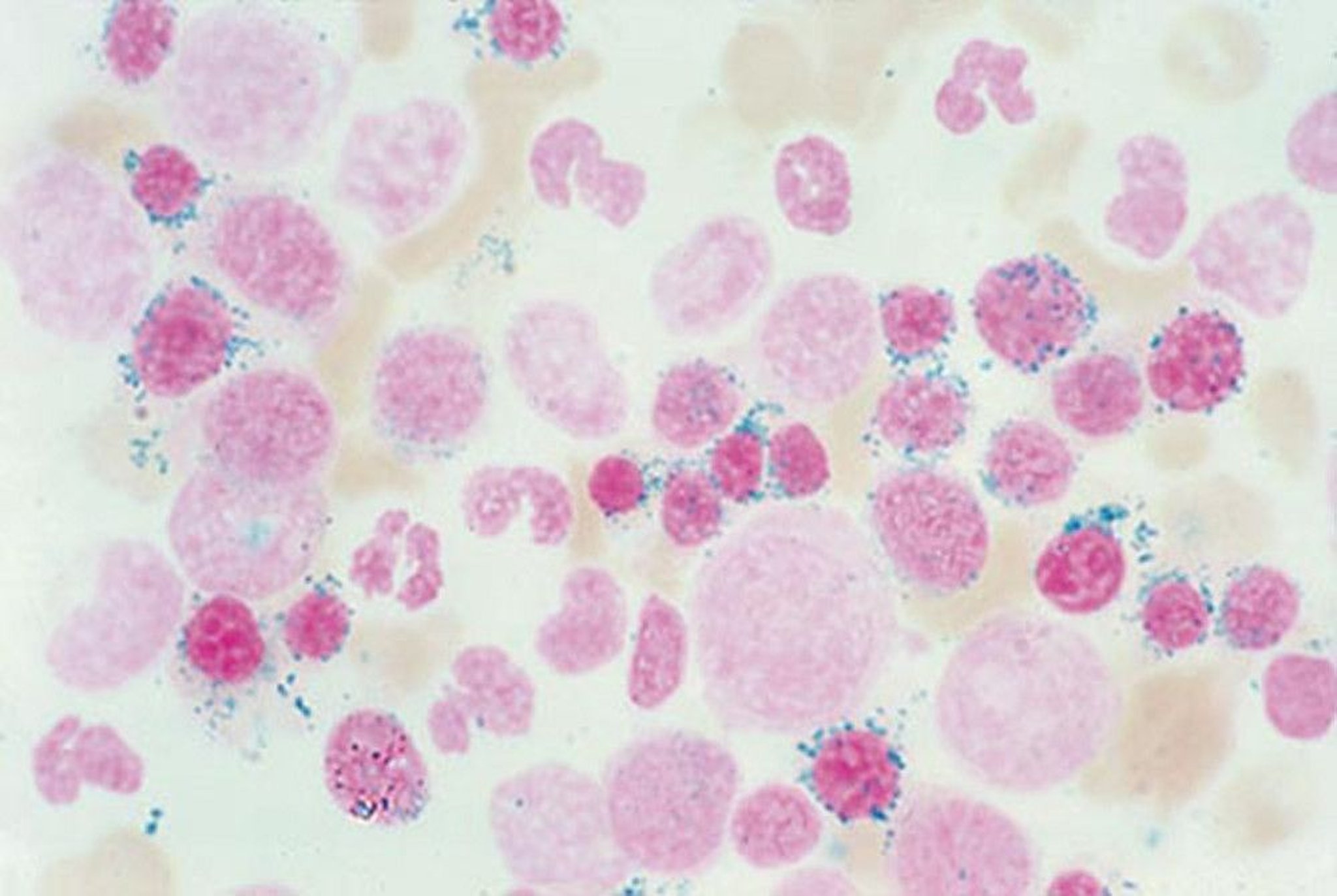
By permission of the publisher. From Tefferi A, Li C. In Atlas of Clinical Hematology. Edited by JO Armitage. Philadelphia, Current Medicine, 2004.
Serum lead is measured if sideroblastic anemia has an unknown cause.
Treatment of Sideroblastic Anemias
Stopping causative substances
Vitamin or mineral supplementation
Recombinant erythropoietin (EPO)
Acquired cases will frequently respond to high doses of recombinant EPO and standard management for low-risk myelodysplastic syndrome1).
Treating iron overload with chelation or phlebotomy as tolerated helps to prevent end organ damage. Management of severe anemia is supportive with transfusions. In young patients with congenital cases who are transfusion dependent, allogeneic bone marrow transplant should be considered.
Treatment reference
1. Fenaux P, Platzbecker U, Mufti GJ, et al:Luspatercept in patients with lower-risk myelodysplastic syndromes. N Engl J Med 382(2): 140–151, 2020. doi: 10.1056/NEJMoa1908892
Key Points
Sideroblastic anemia can be acquired or congenital.
Ringed sideroblasts on a bone marrow biopsy are pathognomic.
Anemia is usually microcytic in congenital sideroblastic anemia and macrocytic in acquired sideroblastic anemia.
Serum iron, ferritin, and transferrin are typically increased.


