Search
SectionsIndexSymptoms
  • Cardiovascular Disorders
  • Clinical Pharmacology
  • Critical Care Medicine
  • Dental Disorders
  • Dermatologic Disorders
  • Ear, Nose, and Throat Disorders
  • Endocrine and Metabolic Disorders
  • Eye Disorders
  • Gastrointestinal Disorders
  • Genitourinary Disorders
  • Geriatrics
  • Gynecology and Obstetrics
  • Hematology and Oncology
  • Hepatic and Biliary Disorders
  • Immunology; Allergic Disorders
  • Infectious Diseases
  • Injuries; Poisoning
  • Musculoskeletal and Connective Tissue Disorders
  • Neurologic Disorders
  • Nutritional Disorders
  • Pediatrics
  • Psychiatric Disorders
  • Pulmonary Disorders
  • Special Subjects
ABCDEFGHI
JKLMNOPQR
STUVWXYZ
  • Abdominal Pain, Acute
  • Abdominal pain, Chronic
  • Alopecia
  • Amenorrhea
  • Amnesia
  • Anosmia
  • Bleeding, Excessive
  • Breast Lumps
  • Chest Pain
  • Constipation in Adults
  • Constipation in Children
  • Cough in Adults
  • Cough in Children
  • Crying
  • Diarrhea in Adults
  • Diarrhea in Children
  • Diplopia
  • Dizziness
  • Dry Mouth
  • Dysmenorrhea
  • Dyspepsia
  • Dysphagia
  • Dyspnea
  • Dysuria
  • Earache
  • Ear Discharge
  • Edema
  • Edema During Late Pregnancy
  • Epistaxis
  • Erectile dysfunction
  • Eyelid Swelling
  • Eye Pain
  • Fever
  • Fever, Acute, in Adults
  • Fever, Chronic (FUO)
  • Fever in Infants and Children
  • Floaters
  • Gas
  • Gastrointestinal Bleeding
  • Halitosis
  • Headache
  • Hearing Loss
  • Hearing Loss: Sudden Deafness
  • Hematospermia
  • Hematuria
  • Hemoptysis
  • Hiccups
  • Hirsutism
  • Insomnia and Excessive Daytime Sleepiness
  • Itching
  • Itching, Anal
  • Jaundice in Adults
  • Jaundice in Neonates
  • Joint Pain, Monarticular
  • Joint Pain, Polyarticular
  • Knee pain
  • Lump in Throat
  • Nasal Congestion and Rhinorrhea
  • Nausea and Vomiting During Early pPregnancy
  • Nausea and Vomiting in Adults
  • Nausea and Vomiting in Infants and Children
  • Neck and Back Pain
  • Neck Mass
  • Nipple Discharge
  • Orthostatis Hypotension
  • Pain
  • Pain, Chronic
  • Palpitations
  • Pelvic Pain
  • Pelvic Pain During Early Pregnancy
  • Polyuria
  • Priapism
  • Red Eye
  • Scrotal Pain
  • Sore Throat
  • Stomatitis
  • Stridor
  • Syncope
  • Tearing
  • Tinnitus
  • Toothache
  • Tremor
  • Urinary Frequency
  • Urinary Incontinence in Adults
  • Urinary Incontinence in Children
  • Urinary Retention
  • Urticaria
  • Vaginal Bleeding
  • Vaginal Bleeding During Early Pregnancy
  • Vaginal Bleeding During Late Pregnancy
  • Vaginal Itching and Discharge
  • Vision, Blurred
  • Vision Loss, Acute
  • Weakness, Generalized
  • Wheezing
In This Topic
Hematology and Oncology
Hemostasis
Overview of Hemostasis
Vascular Factors
Platelet Factors
Plasma Factors
Regulatory Mechanisms
Inactivation of coagulation factors
Fibrinolysis
Regulation of fibrinolysis
Back to Top
Resources
  • About The Merck Manual
  • Ready Reference Guides
  • Trade Names of Some Commonly Used Drugs
  • Normal Laboratory Values
  • Clinical Calculators
  • Multimedia
  • Selected Links
Manuals available online
'/home/index.html' + bookPageLink
 
'/professional/index.html'
These and other Manuals available
in print, online, and as mobile applications.

See more at MerckManuals.com
Sections in Health Care Professionals
  • Cardiovascular Disorders
  • Clinical Pharmacology
  • Critical Care Medicine
  • Dental Disorders
  • Dermatologic Disorders
  • Ear, Nose, and Throat Disorders
  • Endocrine and Metabolic Disorders
  • Eye Disorders
  • Gastrointestinal Disorders
  • Genitourinary Disorders
  • Geriatrics
  • Gynecology and Obstetrics
  • Hematology and Oncology
  • Hepatic and Biliary Disorders
  • Immunology; Allergic Disorders
  • Infectious Diseases
  • Injuries; Poisoning
  • Musculoskeletal and Connective Tissue Disorders
  • Neurologic Disorders
  • Nutritional Disorders
  • Pediatrics
  • Psychiatric Disorders
  • Pulmonary Disorders
  • Special Subjects
Chapters in Hematology and Oncology
  • Approach to the Patient With Anemia
  • Anemias Caused by Deficient Erythropoiesis
  • Anemias Caused by Hemolysis
  • Neutropenia and Lymphocytopenia
  • Thrombocytopenia and Platelet Dysfunction
  • Hemostasis
  • Thrombotic Disorders
  • Coagulation Disorders
  • Bleeding Due to Abnormal Blood Vessels
  • Spleen Disorders
  • Eosinophilic Disorders
  • Histiocytic Syndromes
  • Myeloproliferative Disorders
  • Leukemias
  • Lymphomas
  • Plasma Cell Disorders
  • Iron Overload
  • Transfusion Medicine
  • Overview of Cancer
  • Tumor Immunology
  • Principles of Cancer Therapy
Topics in Hemostasis
  • Overview of Hemostasis
  • Excessive Bleeding
       
      • Merck Manual
      • >
      • Health Care Professionals
      • >
      • Hematology and Oncology
      • >
      • Hemostasis
      • 4
       
      Overview of Hemostasis

      Share This

      Hemostasis, the arrest of bleeding from an injured blood vessel, requires the combined activity of vascular, platelet, and plasma factors. Regulatory mechanisms counterbalance the tendency of clots to form. Hemostatic abnormalities can lead to excessive bleeding or thrombosis.

      Vascular Factors

      Vascular factors reduce blood loss from trauma through local vasoconstriction (an immediate reaction to injury) and compression of injured vessels by extravasation of blood into surrounding tissues. Vessel wall injury triggers the attachment and activation of platelets and production of fibrin; platelets and fibrin combine to form a clot.

      Platelet Factors

      Various mechanisms, including endothelial cell nitric oxide and prostacyclin, promote blood fluidity by preventing platelet stasis and dilating intact blood vessels. These mediators are no longer produced when the vascular endothelium is disrupted. Under these conditions, platelets adhere to the damaged intima and form aggregates. Initial platelet adhesion is to von Willebrand's factor (VWF), previously secreted by endothelial cells into the subendothelium. VWF binds to receptors on the platelet surface membrane (glycoprotein Ib/IX). Platelets anchored to the vessel wall undergo activation. During activation, platelets release mediators from storage granules, including adenosine diphosphate (ADP). Other biochemical changes resulting from activation include hydrolysis of membrane phospholipids, inhibition of adenylate cyclase, mobilization of intracellular Ca, and phosphorylation of intracellular proteins. Arachidonic acid is converted to thromboxane A2; this reaction requires cyclooxygenase and is inhibited irreversibly by aspirinSome Trade Names
      BUFFERIN
      ECOTRIN
      GENACOTE
      Click for Drug Monograph
      and reversibly by many NSAIDs. ADP, thromboxane A2, and other mediators induce activation and aggregation of additional platelets on the injured endothelium. Another receptor is assembled on the platelet surface membrane from glycoproteins IIb and IIIa. Fibrinogen binds to the glycoprotein IIb/IIIa complexes of adjacent platelets, connecting them into aggregates.

      Platelets provide surfaces for the assembly and activation of coagulation complexes and the generation of thrombin. Thrombin converts fibrinogen to fibrin. Fibrin strands bind aggregated platelets to help secure the platelet-fibrin hemostatic plug.

      Plasma Factors

      Plasma coagulation factors interact to produce thrombin, which converts fibrinogen to fibrin. Radiating from and anchoring the hemostatic plug, fibrin strengthens the clot.

      In the intrinsic pathway, factor XII, high molecular weight kininogen, prekallikrein, and activated factor XI (factor XIa) interact to produce factor IXa from factor IX. Factor IXa then combines with factor VIIIa and procoagulant phospholipid (present on the surface of activated platelets and tissue cells) to form a complex that activates factor X. In the extrinsic pathway, factor VIIa and tissue factor directly activate factor X (the factor VIIa/tissue factor complex also activates factor IX—see Fig. 1: Hemostasis: Pathways in blood coagulation.Figures and Table 1: Hemostasis: Components of Blood Coagulation ReactionsTables).

      Fig. 1

      Pathways in blood coagulation.

      Table 1

      PrintOpen table in new window Open table in new window
      Components of Blood Coagulation Reactions

      Factor Number or Name

      Synonym

      Purpose

      Plasma factors

      I

      Fibrinogen

      A precursor of fibrin

      II

      Prothrombin

      A precursor of thrombin, which converts fibrinogen to fibrin; activates factors V, VIII, XI, and XIII; and binds to thrombomodulin to activate protein C

      Is vitamin K–dependent

      V

      Proaccelerin

      Is activated to factor Va, which is a cofactor for the enzyme factor Xa in the factor Xa/Va/phospholipid complex, which cleaves prothrombin to thrombin

      Is present in α granules in platelets

      Factor Va inactivated by activated protein C in complex with free protein S

      VII

      Proconvertin

      Binds to tissue factor and is then activated to form the enzymatic component of the factor VIIa/tissue factor complex, which activates factors IX and X

      Is vitamin K–dependent

      VIII

      Antihemophilic globulin

      Is activated to factor VIIIa, which is a cofactor for the enzyme factor IXa in the factor IXa/VIIIa/phospholipid complex, which activates factor X

      Is a large cofactor protein (as is factor V)

      Circulates in plasma bound to von Willebrand's factor multimers

      As factor VIIIa, is inactivated by activated protein C in complex with free protein S (as is factor Va)

      IX

      Christmas factor

      Is activated to factor IXa, which is the enzyme of the factor IXa/VIIIa/phospholipid complex, which activates factor X

      Is vitamin K–dependent

      X

      Stuart-Prower factor

      Is activated to factor Xa, which is the enzyme of the factor Xa/Va/phospholipid complex, which cleaves prothrombin to thrombin

      Is vitamin K–dependent

      XI

      Plasma thromboplastin antecedent

      Is activated to factor XIa, which activates factor IX in a reaction requiring Ca2+ ions

      Prekallikrein

      Fletcher factor

      Participates in a reciprocal reaction in which it is activated to kallikrein by factor XIIa

      As kallikrein, catalyzes further activation of factor XII to factor XIIa

      Circulates as a biomolecular complex with high molecular weight kininogen

      High molecular weight kininogen

      Fitzgerald factor

      Circulates as a bimolecular complex with prekallikrein

      XII

      Hageman factor

      When activated to factor XIIa by surface contact, kallikrein, or other factors, activates prekallikrein and factor XI, triggering the intrinsic coagulation pathway in vitro

      XIII

      Fibrin stabilizing factor

      When activated by thrombin, catalyzes formation of peptide bonds between adjacent fibrin monomers, thus strengthening and stabilizing the fibrin clot

      Protein C

      —

      Is activated by thrombin bound to thrombomodulin; then proteolyzes and inhibits (in the presence of free protein S and phospholipid) the cofactor activity of factors VIIIa and Va

      Is vitamin K–dependent

      Protein S

      —

      Circulates in plasma as free protein S and as protein S bound to C4b-binding protein of the complement system

      Functions in its free form as a cofactor for activated protein C

      Is vitamin K–dependent

      Cell surface factors

      Tissue factor

      Tissue thromboplastin

      Is a lipoprotein that is constitutively present on the membrane of certain tissue cells, including perivascular fibroblasts, boundary epithelial cells (eg, epithelial cells of the skin, amnion, and GI and GU tracts), and glial cells of the nervous system

      May also develop in pathologic states on activated monocytes and macrophages and on activated vascular endothelium

      Is present on some tumor cells

      Binds factor VIIa, which initiates the extrinsic coagulation pathway

      Procoagulant phospholipid

      —

      Acidic phospholipid (primarily phosphatidyl serine) present on the surface of activated platelets and other tissue cells

      Is a component of the factor IXa/VIIIa/phospholipid complex which activates factor X and of the factor Xa/Va/phospholipid complex which activates prothrombin

      Functions as the lipid moiety of tissue factor

      Thrombomodulin

      —

      Is an endothelial cell surface binding site for thrombin, which, when bound to thrombomodulin, activates protein C

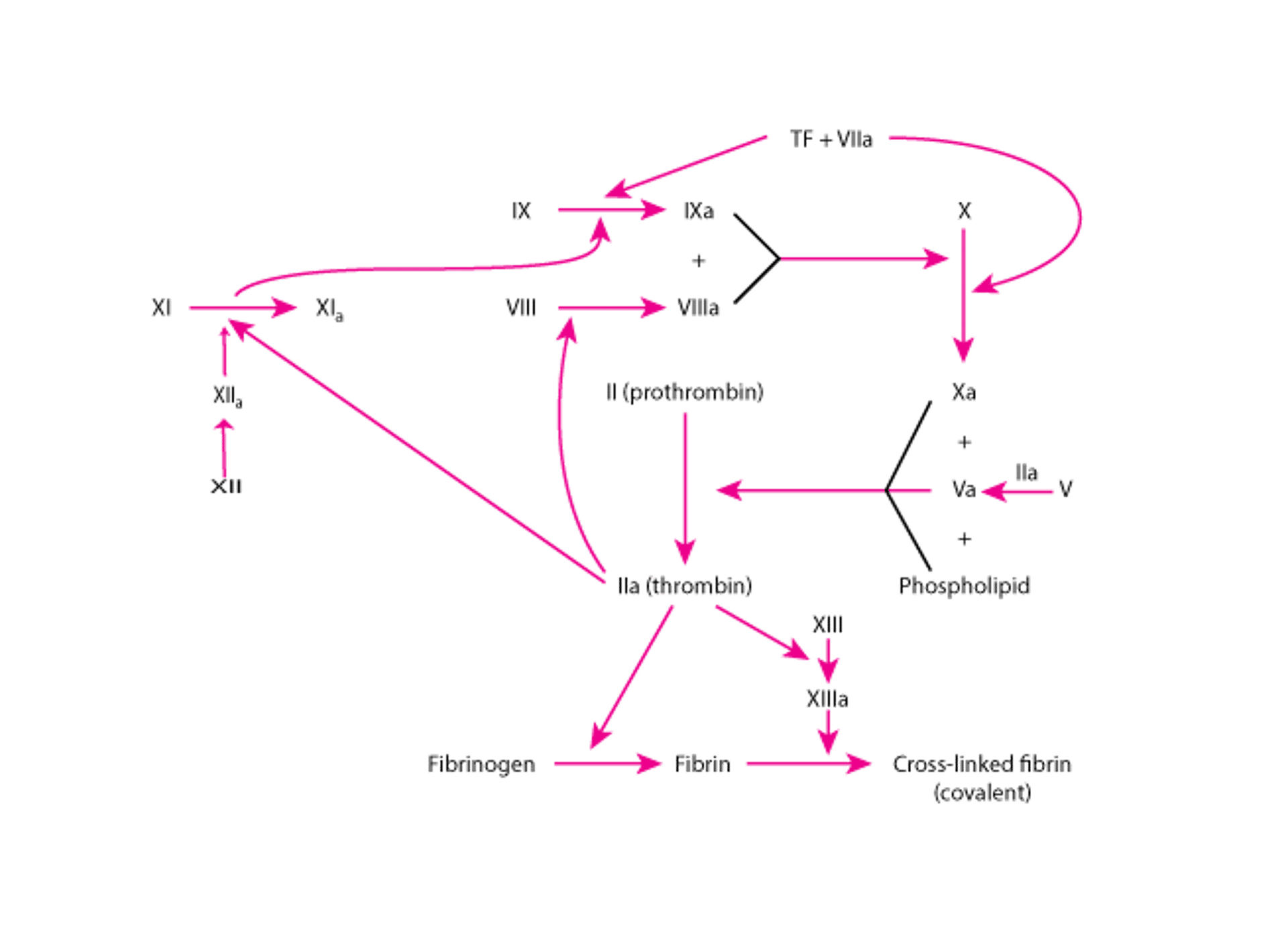
      Activation of the intrinsic or extrinsic pathway activates the common pathway, resulting in formation of the fibrin clot. Three steps are involved in common pathway activation:

      1. A prothrombin activator is produced on the surface of activated platelets and tissue cells. The activator is a complex of an enzyme, factor Xa, and 2 cofactors, factor Va and procoagulant phospholipid.
      2. The prothrombin activator cleaves prothrombin into thrombin and another fragment.
      3. Thrombin induces the generation of fibrin polymers from fibrinogen. Thrombin also activates factor XIII, an enzyme that catalyzes formation of stronger bonds between adjacent fibrin monomers, as well as factor VIII and factor XI.

      Ca2+ ions are needed in most thrombin-generating reactions (Ca2+-chelating agents [eg, citrate, ethylenediaminetetraacetic acid] are used in vitro as anticoagulants). Vitamin K–dependent clotting factors (factors II, VII, IX, and X) cannot bind normally to phospholipid surfaces through Ca2+ bridges and function in blood coagulation when the factors are synthesized in the absence of vitamin K.

      Although the coagulation pathways are helpful in understanding mechanisms and laboratory evaluation of coagulation disorders, in vivo coagulation is predominantly via the extrinsic pathway. People with hereditary deficiencies of factor XII, high molecular weight kininogen, or prekallikrein have no bleeding abnormality. People with hereditary factor XI deficiency have a mild to moderate bleeding disorder. In vivo, factor XI (an intrinsic pathway factor) is activated when a small amount of thrombin is generated. Factor IX can be activated both by factor XIa and factor VIIa/tissue factor complexes.

      In vivo, initiation of the extrinsic pathway occurs when injury to blood vessels brings blood into contact with tissue factor on membranes of cells within and around the vessel walls. This contact with tissue factor generates factor VIIa/tissue factor complexes that activate factor X and factor IX. Factor IXa, combined with its cofactor, factor VIIIa, on phospholipid membrane surfaces generates additional factor Xa. Factor X activation by both factor VIIa/tissue factor and factor IXa/VIIIa complexes is required for normal hemostasis. This requirement for factors VIII and IX explains why hemophilia type A (deficiency of factor VIII) or type B (deficiency of factor IX) results in bleeding despite an intact extrinsic coagulation pathway initiated by factor VIIa/tissue factor complexes.

      Regulatory Mechanisms

      Several inhibitory mechanisms prevent activated coagulation reactions from amplifying uncontrollably, causing extensive local thrombosis or disseminated intravascular coagulation. These mechanisms include

      • Inactivation of procoagulant enzymes
      • Fibrinolysis
      • Hepatic clearance of activated clotting factors

      Inactivation of coagulation factors: Plasma protease inhibitors (antithrombin, tissue factor pathway inhibitor, α2-macroglobulin, heparin cofactor II) inactivate coagulation enzymes. Antithrombin inhibits thrombin, factor Xa, factor XIa, and factor IXa. Heparin enhances antithrombin activity.

      Two vitamin K–dependent proteins, protein C and free protein S, form a complex that inactivates factors VIIIa and Va by proteolysis. Thrombin, when bound to a receptor on endothelial cells (thrombomodulin), activates protein C. Activated protein C, in combination with free protein S and phospholipid cofactors, proteolyzes and inactivates factors VIIIa and Va.
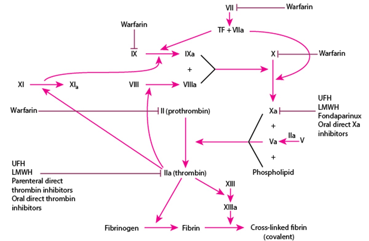
      Fibrinolysis: Fibrin deposition and lysis must be balanced to maintain temporarily and subsequently remove the hemostatic seal during repair of an injured vessel wall. The fibrinolytic system dissolves fibrin by means of plasmin, a proteolytic enzyme. Fibrinolysis is activated by plasminogen activators released from vascular endothelial cells. Plasminogen activators and plasminogen (from plasma) bind to fibrin, and plasminogen activators cleave plasminogen into plasmin (see Fig. 2: Hemostasis: Fibrinolytic pathway.Figures). Plasmin then proteolyzes fibrin into soluble fibrin degradation products that are swept away in the circulation.

      Fig. 2

      Fibrinolytic pathway.

      Fibrin deposition and fibrinolysis must be balanced during repair of an injured blood vessel wall. Injured vascular endothelial cells release plasminogen activators (tissue plasminogen activator, urokinase), activating fibrinolysis. Plasminogen activators cleave plasminogen into plasmin, which dissolves clots. Fibrinolysis is controlled by plasminogen activator inhibitors (PAIs; eg, PAI-1) and plasmin inhibitors (eg, α2-antiplasmin).

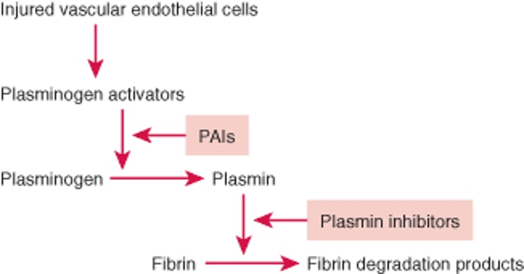
      There are several plasminogen activators:

      • Tissue plasminogen activator (tPA), from endothelial cells, is a poor activator when free in solution but an efficient activator when bound to fibrin in proximity to plasminogen.
      • Urokinase exists in single-chain and double-chain forms with different functional properties. Single-chain urokinase cannot activate free plasminogen but, like tPA, can readily activate plasminogen bound to fibrin. A trace concentration of plasmin cleaves single-chain to double-chain urokinase, which activates plasminogen in solution as well as plasminogen bound to fibrin. Epithelial cells that line excretory passages (eg, renal tubules, mammary ducts) secrete urokinase, which is the physiologic activator of fibrinolysis in these channels.
      • Streptokinase, a bacterial product not normally found in the body, is another potent plasminogen activator.

      StreptokinaseSome Trade Names
      STREPTASE

      , urokinase, and recombinant tPA (alteplaseSome Trade Names
      ACTIVASE
      Click for Drug Monograph
      ) have all been used therapeutically to induce fibrinolysis in patients with acute thrombotic disorders.

      Regulation of fibrinolysis: Fibrinolysis is regulated by plasminogen activator inhibitors (PAIs) and plasmin inhibitors that slow fibrinolysis. PAI-1, the most important PAI, inactivates tPA and urokinase and is released from vascular endothelial cells and activated platelets. The primary plasmin inhibitor is α2-antiplasmin, which quickly inactivates any free plasmin escaping from clots. Some α2-antiplasmin is also cross-linked to fibrin polymers by the action of factor XIIIa during clotting. This cross-linking may prevent excessive plasmin activity within clots.

      tPA and urokinase are rapidly cleared by the liver, which is another mechanism of preventing excessive fibrinolysis.

      Last full review/revision July 2012 by Joel L. Moake, MD

      Content last modified July 2012

      Buy the Book

      Mobile Versions

      Back to Top

      Previous: Overview of Platelet Disorders

      Next: Excessive Bleeding

      Audio
      Figures
      Photographs
      Sidebars
      Tables
      Videos

      Copyright     © 2010-2013 Merck Sharp & Dohme Corp., a subsidiary of Merck & Co., Inc., Whitehouse Station, N.J., U.S.A.    Privacy    Terms of Use