


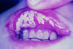
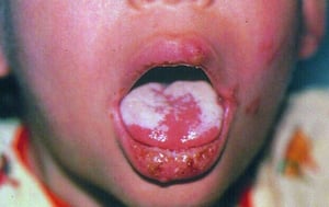
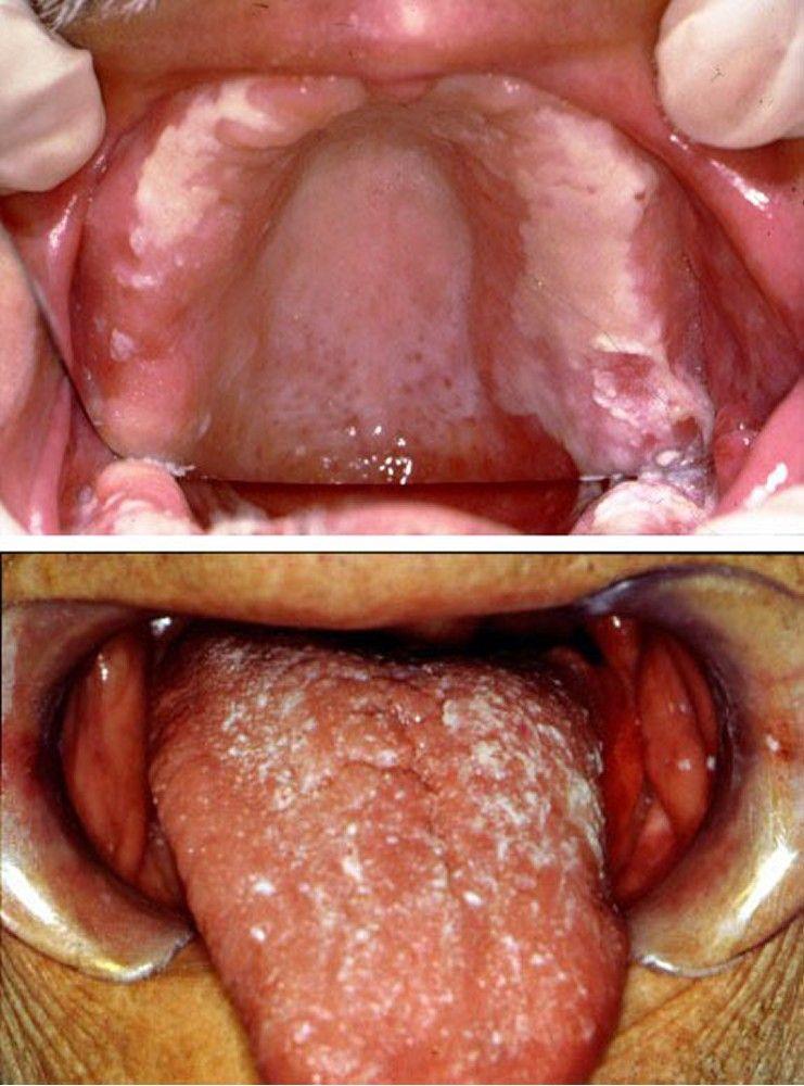
Candidiasis is infection by Candida species (most often C. albicans), manifested by mucocutaneous lesions, fungemia, and sometimes focal infection of multiple sites. Symptoms depend on the site of infection and include dysphagia, skin and mucosal lesions, blindness, vulvovaginal symptoms (itching, burning, discharge), fever, shock, oliguria, renal shutdown, and disseminated intravascular coagulation. Diagnosis is confirmed by cultures from normally sterile sites. For candidemia and invasive candidiasis, initial treatment with an echinocandin and potential step-down to fluconazole or another azole is recommended.
(See also Overview of Fungal Infections, Candidiasis (Mucocutaneous), Candidal Vaginitis, and Chronic Mucocutaneous Candidiasis.)
Candida species are commensal organisms that inhabit the gastrointestinal (GI) tract and sometimes the skin (see etiology of mucocutaneous candidiasis). Unlike other systemic mycoses, candidiasis results from endogenous organisms.
Most infections are caused by C. albicans; however, Nakaseomyces glabrata (C. glabrata) and other non-albicans species are increasingly involved in fungemia, urinary tract infections, and, occasionally, other focal disease.
Antimicrobial sensitivities vary across Candida species:
N. glabrata
Pichia kudriavzevii (C. krusei) is inherently resistant to fluconazole; frequency of resistance to voriconazole and amphotericin B varies. P. kudriavzevii is most frequently susceptible to echinocandins.
C. auris is an emerging, multidrug-resistant species that has caused outbreaks in hospitals.
Candida species are a major cause of systemic fungal infections and are the most common cause of fungal infections in patients who are immunocompromised. Candidal infections are one of the most common hospital-acquired infections. Because resistance and transmission of C. auris in health care facilities have become a concern, special infection control precautions have been instituted for patients who are colonized or infected with C. auris.
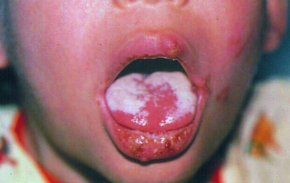
Candidiasis of the esophagus is a defining opportunistic infection in AIDS. Although mucocutaneous candidiasis is frequently present in patients with AIDS, hematogenous dissemination is unusual unless other specific risk factors are present (see Disseminated candidiasis).


Images courtesy of Jonathan Ship, MD.
© Springer Science+Business Media
© Springer Science+Business Media

Images courtesy of Jonathan Ship, MD.


© Springer Science+Business Media

© Springer Science+Business Media

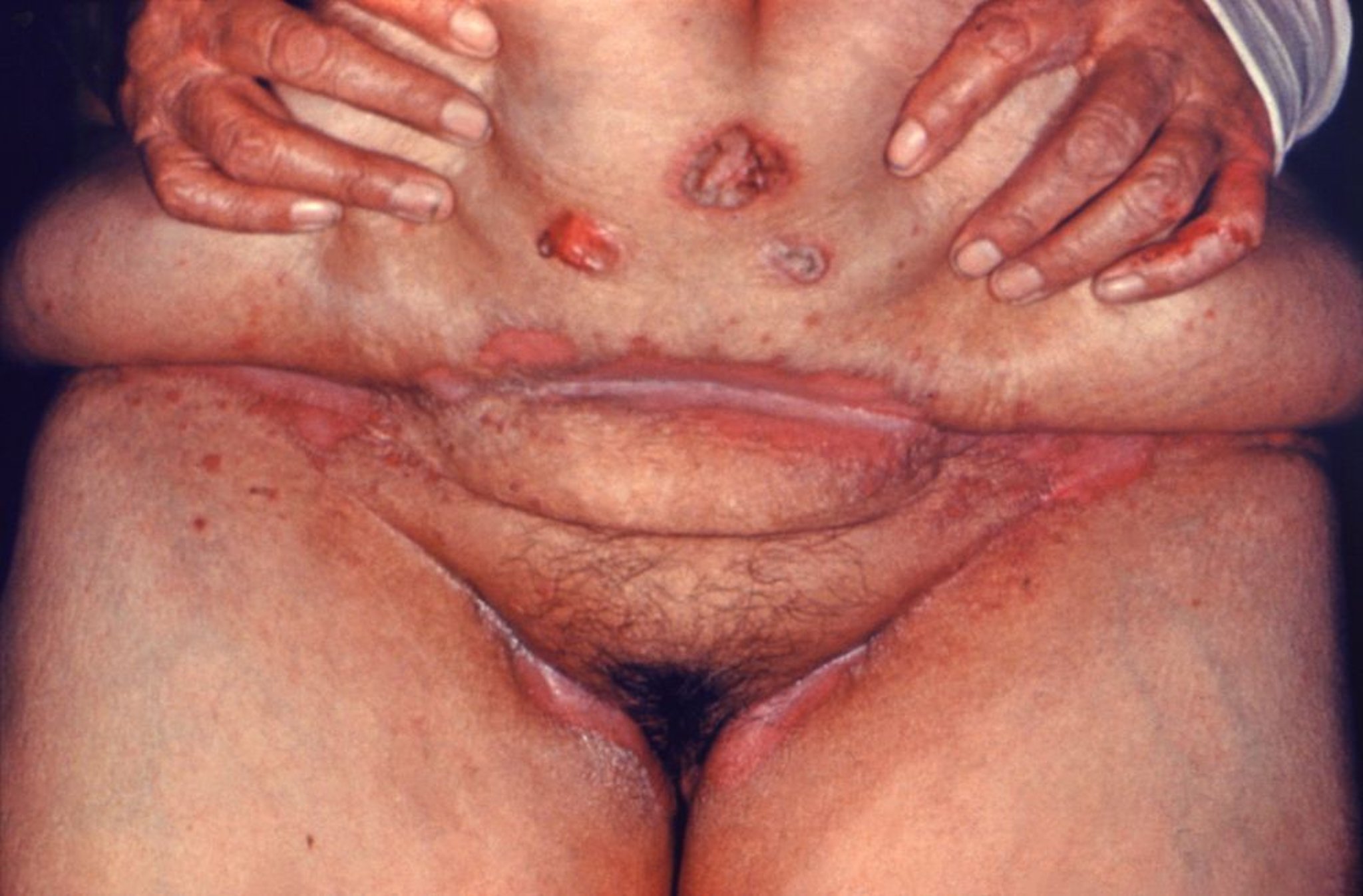
Image courtesy of Dr. Hardin via the Public Health Image Library of the Centers for Disease Control and Prevention.
Disseminated candidiasis
Neutropenic patients (eg, during cancer chemotherapy) are at high risk of developing life-threatening disseminated candidiasis.
Candidemia may occur in nonneutropenic patients during prolonged hospitalization. This bloodstream infection is often related to one or more of the following:
Central venous catheters
Major surgery
Broad-spectrum antibacterial therapy
IV hyperalimentation
IV lines and the GI tract are the usual portals of entry.
Candidemia often prolongs hospitalization and increases mortality due to concurrent disorders. Candidemia may occur with other forms of invasive candidiasis, such as endocarditis or meningitis, as well as focal involvement of skin, subcutaneous tissues, bones, joints, liver, spleen, kidneys, eyes, and other tissues. Endocarditis is commonly related to IV use of illicit drugs, valve replacement, or intravascular trauma induced by indwelling IV catheters.
All forms of disseminated candidiasis should be considered serious, progressive, and potentially fatal.
Symptoms and Signs of Invasive Candidiasis
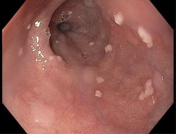
Esophageal candidiasis is most often manifested by dysphagia.
Candidemia usually causes fever, but symptoms are nonspecific. Some patients develop a syndrome resembling bacterial sepsis, with a fulminating course that may include shock, oliguria, acute kidney injury, and disseminated intravascular coagulation.
Candidal endophthalmitis starts as white retinal lesions that are initially asymptomatic but can progress, opacifying the vitreous and causing potentially irreversible scarring and blindness. In neutropenic patients, retinal hemorrhages occasionally occur, but actual infection of the eye is rare.
Papulonodular skin lesions may also develop, especially in neutropenic patients, in whom they indicate widespread hematogenous dissemination to other organs. Symptoms of other focal or invasive infection depend on the organ involved.
Diagnosis of Invasive Candidiasis
Histopathology and fungal cultures
Blood cultures
Serum (1,3)beta-D-glucan
T2Candida Panel
Because Candida species are commensal, their culture from sputum, the mouth, the vagina, urine, stool, or skin does not necessarily signify an invasive, progressive infection. A characteristic clinical lesion must also be present, histopathologic evidence of tissue invasion (eg, yeasts, pseudohyphae, or hyphae in tissue specimens) must be documented, and other etiologies must be excluded. Positive cultures of specimens taken from normally sterile sites, such as blood, cerebrospinal fluid, pericardium, pericardial fluid, or biopsied tissue, provide definitive evidence that systemic therapy is needed.
Standard laboratory techniques often misidentify C. auris as C. haemulonii, C. famata, C. sake, or another species. MALDI-TOF mass spectrometry is a more reliable method for correct identification. A nucleic acid-based test also is now available.
Serum (1,3)beta-D-glucan is often positive in patients with invasive candidiasis; conversely, a negative result indicates low likelihood of systemic infection.
The T2Candida Panel is a magnetic resonance assay that directly detects 5 Candida species (C. albicans, C. tropicalis, C. parapsilosis, P. kudriavzevii, and N. glabrata) in whole blood samples in 3 to 5 hours. It is highly sensitive and has an excellent negative predictive value (1). Other molecular diagnostic testing is also available, including matrix-assisted laser desorption ionization–time of flight (MALDI-TOF) mass spectrometry and polymerase chain reaction (PCR)-based assays.
Ophthalmologic examination to check for endophthalmitis is recommended for patients with candidemia. Experts' opinions vary regarding whether fundoscopic examination is required for all patients or only those with visual symptoms.

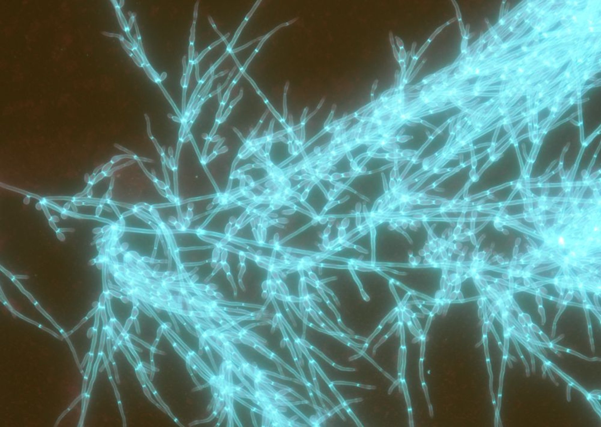
Image courtesy of Paschalis Vergidis, MD, MSc.
Diagnosis reference
1. Zervou FN, Zacharioudakis IM, Kurpewski J, Mylonakis E: T2 magnetic resonance for fungal diagnosis. Methods Mol Biol 1508:305–319, 2017. doi: 10.1007/978-1-4939-6515-1_18
Treatment of Invasive Candidiasis
An echinocandin if patients are severely or critically ill or if infection with N. glabrata, C. auris, or P. kudriavzevii is suspected
C. albicans or C. parapsilosis is suspected, or once antifungal susceptibilities become available
Alternatively voriconazole or amphotericin B
(See also Antifungal Medications.)
For candidemia and invasive candidiasis, initial treatment with an echinocandin and potential step-down to fluconazole or another azole is recommended.
Invasive candidiasis
In patients with invasive candidiasis, predisposing conditions (eg, neutropenia, immunosuppression, use of broad-spectrum antibacterial antibiotics, hyperalimentation, presence of indwelling lines) should be reversed or controlled if possible.
In nonneutropenic patients, IV catheters should be removed.
When an echinocandin is indicated (if patients are moderately severely ill or critically ill [most neutropenic patients] or if N. glabrata, C. auris, or P. kudriavzevii is suspected), one of the following medications can be used:
C. albicans or C. parapsilosis is suspected, or once antifungal susceptibilities become available.
1).
Treatment of candidemia is continued for 14 days after the last negative blood culture.
Esophageal candidiasis
Esophageal candidiasis is treated with one of the following:
If these medications are ineffective or if infection is severe, one of the following may be used:
An echinocandin
Treatment of esophageal candidiasis is continued for 14 to 21 days.
Treatment reference
1. Pappas PG, Kauffman CA, Andes DR, et al: Clinical practice guideline for the management of candidiasis: 2016 update by the Infectious Diseases Society of America. Clin Infect Dis 62(4):e1–e50, 2016. doi: 10.1093/cid/civ933
Key Points
Unlike other fungal infections, invasive candidiasis is usually due to endogenous organisms.
Invasive infection typically occurs in patients who are immunocompromised and/or who are hospitalized, particularly those who have had surgery or been given broad-spectrum antibiotics.
Positive cultures of specimens taken from normally sterile sites (eg, blood, cerebrospinal fluid, tissue biopsy specimens) are needed to distinguish invasive infection from normal colonization; serum (1,3)beta-D-glucan is often positive in patients with invasive candidiasis.
A T2Candida Panel on whole blood can be used to diagnose a Candida blood infection.
Use an echinocandin if patients are severely or critically ill or if infection with N. glabrata, C. auris, or P. kudriavzevii (C. krusei) is suspected.
C. albicans or C. parapsilosis is suspected, or once antifungal susceptibilities become available.
More Information
The following English-language resources may be useful. Please note that THE MANUAL is not responsible for the content of these resources.
Centers for Disease Control and Prevention (CDC): Infection Prevention and Control for Candida auris
Infectious Diseases Society of America: Clinical Practice Guideline for the Management of Candidiasis: 2016 Update


