


Chromoblastomycosis is a specific type of cutaneous infection caused by one of several species of dematiaceous (pigmented) fungi. Symptoms are ulcerating nodules on exposed body parts. Diagnosis is by appearance, histopathology, and culture. Treatment is with itraconazole, another mold-active azole, or terbinafine and surgical excision.
(See also Overview of Fungal Infections.)
Chromoblastomycosis is a cutaneous infection affecting immunocompetent people, mostly in tropical or subtropical areas; it is characterized by formation of papillomatous nodules that tend to ulcerate.
Chromoblastomycosis often occurs at the site of penetrating injury, particularly in farmers and other agricultural workers without adequate protective footwear and clothing.
Chromoblastomycosis is caused by dark brown or black fungi that produce sclerotic bodies in tissue.
Symptoms and Signs of Chromoblastomycosis
Usually, chromoblastomycosis begins on the foot or leg, but other exposed body parts may be infected, especially where the skin is broken.
Early small, itchy, enlarging papules may resemble dermatophytosis (ringworm). These papules extend to form dull red or violaceous, sharply demarcated patches with indurated bases. Several weeks or months later, new lesions, projecting 1 to 2 mm above the skin, may appear along paths of lymphatic drainage. Hard, dull red or grayish cauliflower-shaped nodular projections may develop in the center of patches and, if the infection is untreated, gradually extend to cover extremities over the course of many years.
Lymphatics may be obstructed, itching may persist, and secondary bacterial superinfections may develop, causing ulcerations and occasionally septicemia.
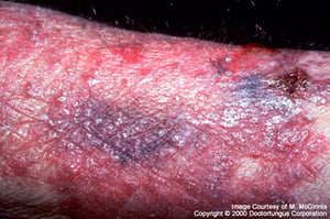
Image courtesy of www.doctorfungus.org © 2005.

Image courtesy of www.doctorfungus.org © 2005.

Image courtesy of Karen McKoy, MD.

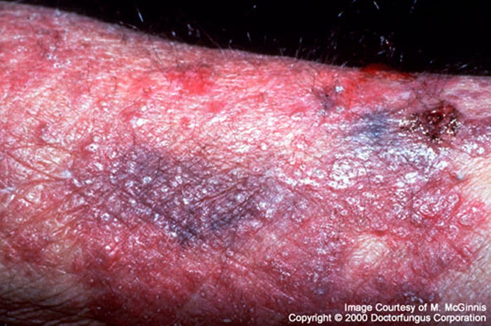
Image courtesy of www.doctorfungus.org © 2005.

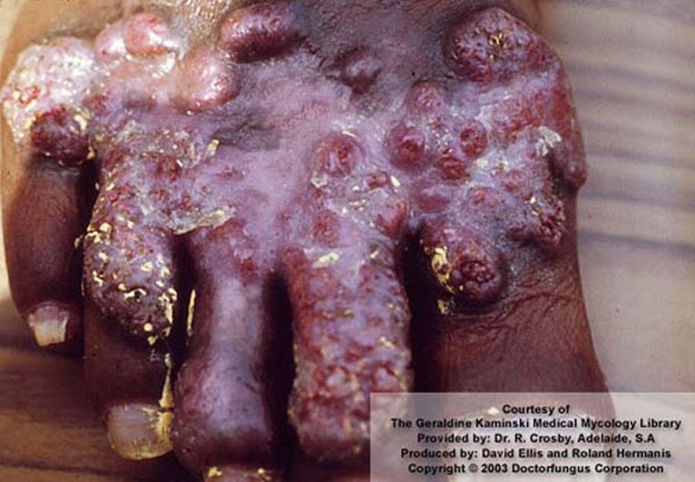
Image courtesy of www.doctorfungus.org © 2005.

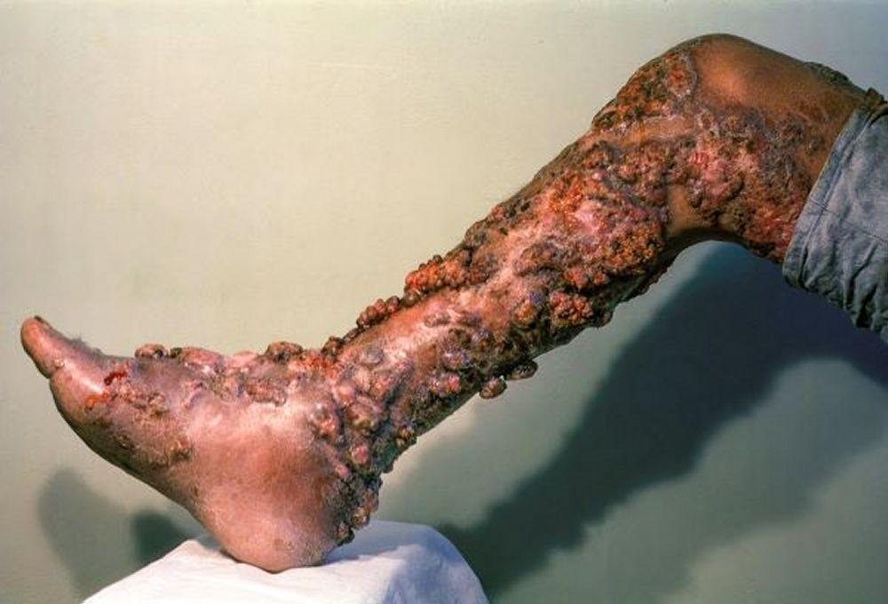
Image courtesy of Karen McKoy, MD.
Diagnosis of Chromoblastomycosis
Histopathology
Culture
Late chromoblastomycosis lesions have a characteristic appearance, but early lesions may be mistaken for dermatophytoses.
Fontana-Masson staining for melanin helps confirm the presence of the sclerotic bodies (Medlar bodies), which are pathognomonic.
Culture is needed to identify the causative species.
Treatment of Chromoblastomycosis
Itraconazole
Terbinafine
Often surgery or cryotherapy
(See also Antifungal Medications.)
Flucytosine should not be used as monotherapy.
For localized lesions, surgical excision may be curative.
Adjunctive therapies such as cryotherapy are often helpful, although response is slow.


