


Most finger dislocations occur at the proximal interphalangeal (PIP) joint; they are usually caused by hyperextension and thus are usually dorsal.
Finger dislocations can be dorsal, lateral, or volar. They may rupture various combinations of supporting ligaments. Most cause obvious deformities, as well as pain and swelling.
Anteroposterior, lateral, and oblique x-rays are taken. Lateral views should be taken with the affected digit visibly separated from the others.
For most dislocations, closed reduction is done after a digital nerve block is used. For all PIP dislocations, stability of the lateral ligaments is assessed by stress testing after the dislocation is reduced.
(See Overview of Dislocations.)
Dorsal dislocations
Dorsal dislocations result from hyperextension. They occasionally displace the volar joint structures intra-articularly (volar plate injury).
In volar plate injuries, x-rays occasionally show a small bone fragment avulsed from the middle phalanx.
Dorsal dislocations are reduced using axial traction and volar force. If volar plate injury is suspected or if closed reduction is difficult (suggesting volar plate injury), open reduction may be necessary.

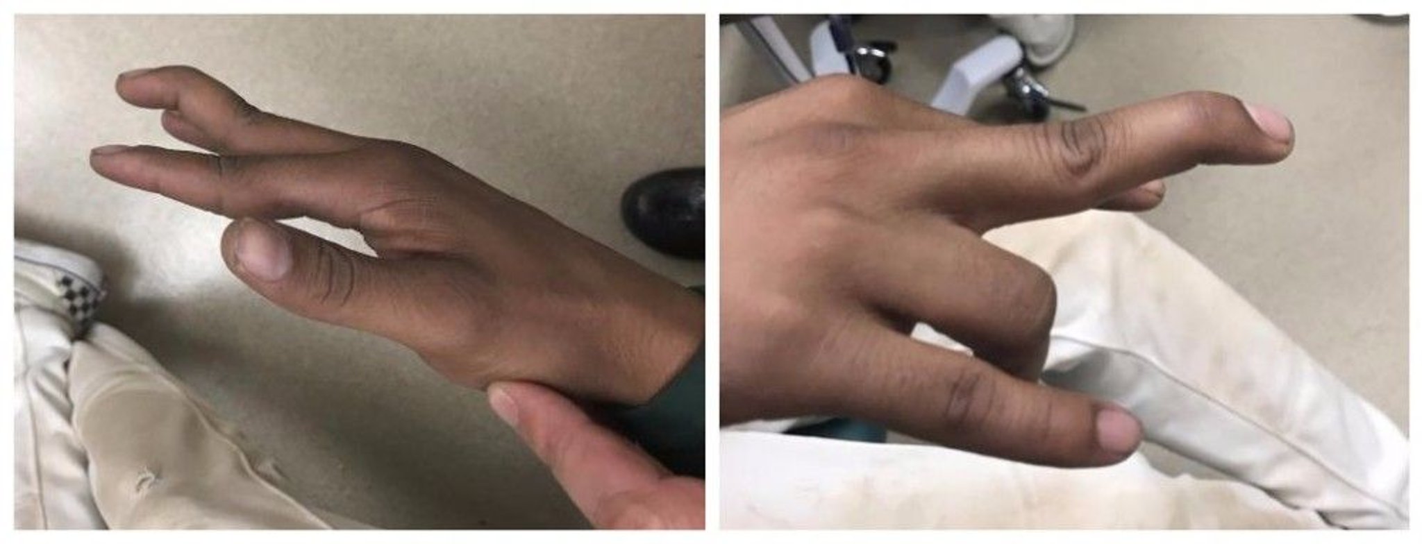
Image courtesy of Danielle Campagne, MD.
Dorsal dislocations are usually splinted in 15° of flexion for 3 weeks.
Lateral dislocations
Lateral dislocations may occur when abduction or adduction forces are applied to an extended finger joint.
The joint is tender and unstable when lateral stress is applied.
The joint is reduced, then splinted in 35° of flexion.
Volar dislocations
Volar dislocations are uncommon and occur when volar forces are applied to a rotated finger joint.

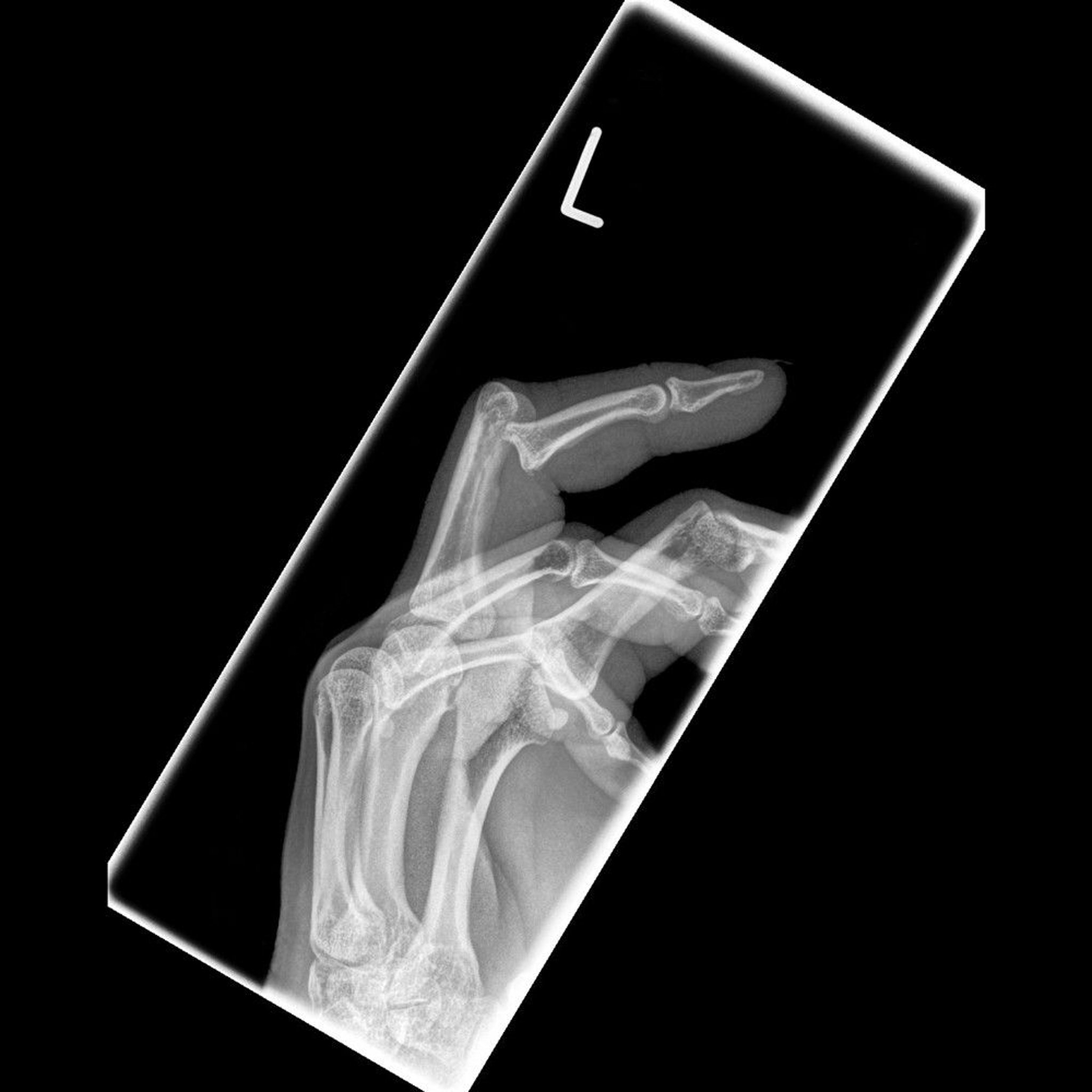
SCIENCE PHOTO LIBRARY
Usually, the central slip of the extensor tendon ruptures, causing boutonnière deformity.
Volar dislocations are reduced using axial traction and dorsal force, then splinted in extension for 1 to 2 weeks. Subsequently, patients should be evaluated to determine whether surgery is needed to repair a ruptured central slip of the extensor tendon.
Key Points
Finger dislocations (dorsal, lateral, or volar) may rupture various combinations of supporting ligaments.
Deformities are usually obvious.
Diagnose by taking anteroposterior, lateral, and oblique x-rays: for lateral views, separate the affected digit from the others.
Reduce most dislocations manually after using a digital nerve block.
After reduction, do stress testing to assess the stability of all PIP dislocations.


