


Lupus Nephritis—Mesangial Proliferative (Class II)

Lupus Nephritis—Mesangial Proliferative (Class II)
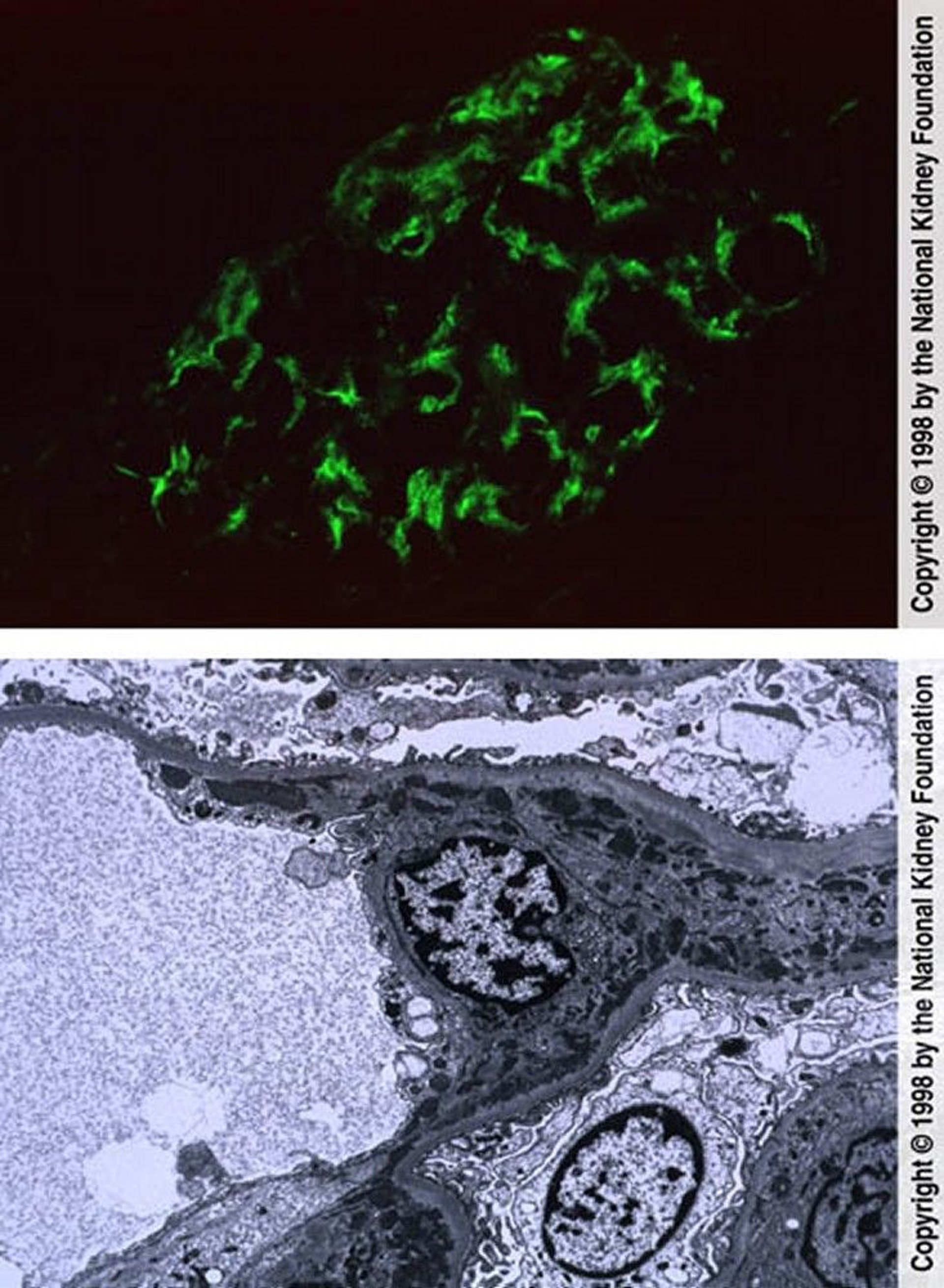
The top image shows mesangial deposition of IgM (immunofluorescence with anti-IgM, original magnification ×400). In the bottom image, mesangial dense immune complex deposits and reticular aggregates within endothelial cell cytoplasm are seen on transmission electron microscopy (×8000).
Image provided by Agnes Fogo, MD, and the American Journal of Kidney Diseases' Atlas of Renal Pathology (see www.ajkd.org).
In these topics