


Cervicothoracic Syringohydromyelia

Cervicothoracic Syringohydromyelia
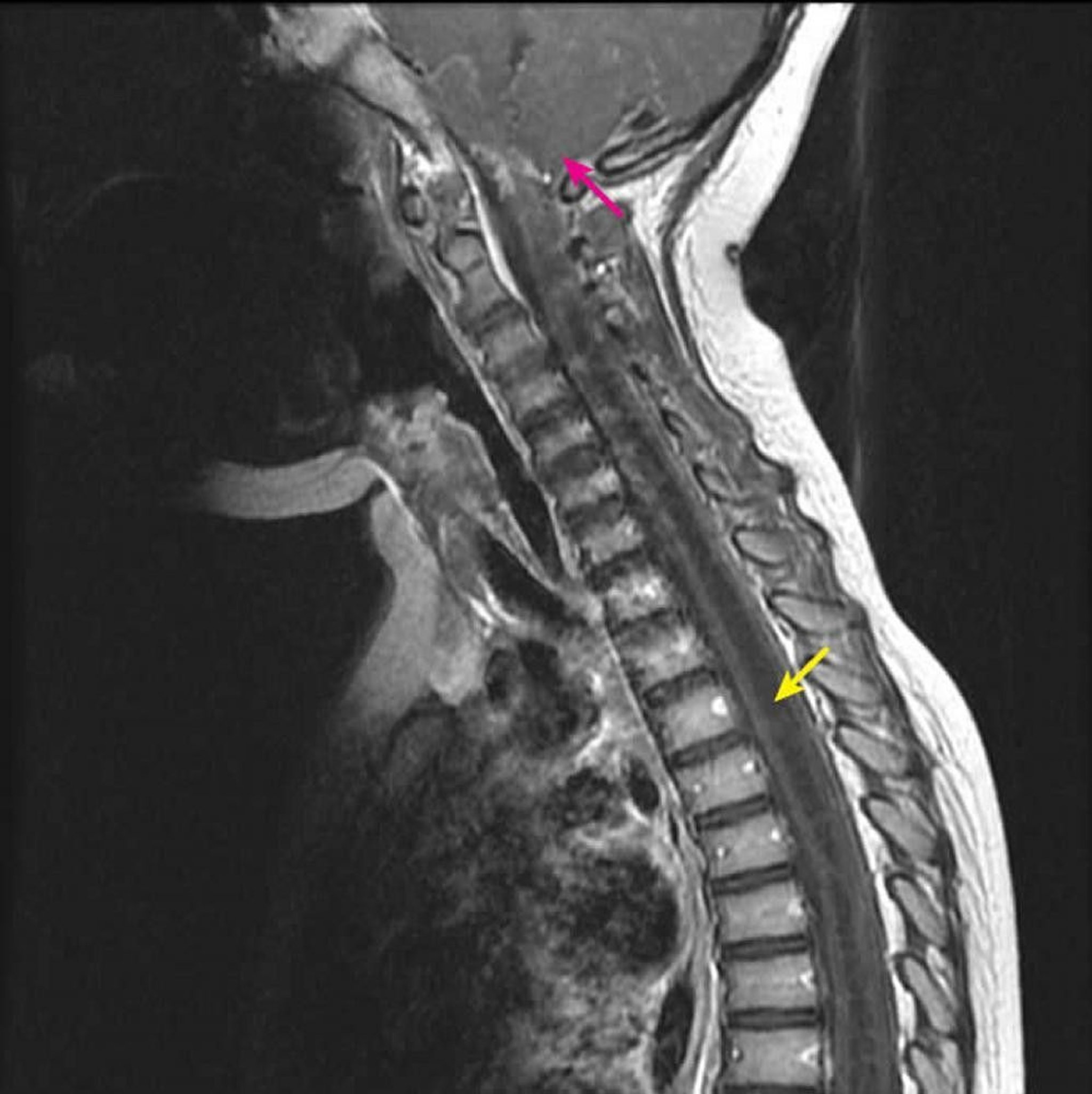
Sagittal post-contrast T1-weighted MRI shows a large T1 hypointense intramedullary spinal cord cavity characteristic of a syrinx (yellow arrow). It extends from the C2-C3 cervical level inferiorly in a child with a known Chiari I malformation (pink arrow). This finding is defined as syringohydromyelia because syringomyelia (an eccentric spinal cord cavity) cannot be differentiated from hydromyelia (a central spinal cord cavity) by imaging.
Courtesy of John Tsiouris, MD, Division of Neuroradiology, New York–Presbyterian Hospital/Weill Cornell Medical Center.
In these topics