


Neurofibromatosis refers to several related genetic disorders that have overlapping clinical manifestations. It causes various types of benign or malignant tumors that involve central or peripheral nerves and often causes pigmented skin macules and sometimes other manifestations. Diagnosis is primarily clinical based on specific criteria. Benign tumors can be removed surgically, and malignant tumors (which are less common) can be treated with radiation therapy or chemotherapy.
Neurofibromatosis is a neurocutaneous syndrome (a syndrome with neurologic and cutaneous manifestations).
Types of Neurofibromatosis
There are 3 main types of neurofibromatosis: neurofibromatosis type 1, NF2-related schwannomatosis (NF2), and non-NF2 schwannomatosis (schwannomatosis). Note that NF2-related schwannomatosis and non-NF2 schwannomatosis are terminologies recommended by the International Consensus Group on Neurofibromatosis Diagnostic Criteria (I-NF-DC) because they better reflect the updated criteria for these disorders that incorporate clinical features and genetic testing (1). (See also table Diagnosing Neurofibromatosis.)
Neurofibromatosis type 1
Neurofibromatosis type 1 (NF1, or von Recklinghausen disease) is most prevalent, occurring in 1 of 4650 people in a study done in the United Kingdom (2). It causes neurologic, cutaneous, and sometimes soft-tissue or bone manifestations. The NF1 gene is located on band 17q11.2 and encodes synthesis of neurofibromin; > 1000 mutations have been identified. Although it is an autosomal dominant disorder, 20 to 50% of cases are caused by a de novo germ cell mutation.
NF2-related schwannomatosis (NF2)
NF2-related schwannomatosis (NF2) accounts for 10% of cases, occurring in about 1 of 25,000 people in a study from the North West of England (3). It manifests primarily as congenital bilateral vestibular schwannoma (acoustic neuroma). The NF2 gene is located on band 22q11 and encodes synthesis of merlin, a tumor suppressor; 200 mutations have been identified. Approximately half of patients with NF2 inherit a mutation from an affected parent (4).
Non-NF2 schwannomatosis
Non-NF2 schwannomatosis (schwannomatosis) is a rare disorder. It is classified as a third type of neurofibromatosis. In 15% of cases, this type is familial and related to a germline mutation in the SMARCB1 or LZTR1 gene. These genes are tumor-suppressor genes and both are located on chromosome 22 very close to the NF2 gene (1). In the remaining cases, the genetic basis is not well-understood, but in tissue from some patients, other mutations in the same gene are involved. Two or more schwannomas develop in spinal and peripheral nerves and are sometimes quite painful; however, vestibular schwannomas do not develop. Schwannomatosis used to be considered a form of NF2 because multiple schwannomas are present in both conditions; however, the clinical picture is different, and the genes involved are distinct.
Types of tumors
Tumors may be peripheral or central.
Peripheral tumors are common in NF1 and can develop anywhere along the course of peripheral nerves. The tumors are neurofibromas, which develop from nerve sheaths and consist of mixtures of Schwann cells, fibroblasts, neural cells, and mast cells. Most appear during adolescence. Occasionally, they transform to malignant peripheral nerve sheath tumors. There are multiple forms:
Cutaneous neurofibromas are soft and fleshy.
Subcutaneous neurofibromas are firm and nodular.
Nodular plexiform neurofibromas may involve spinal nerve roots, typically growing through an intervertebral foramen to cause intraspinal and extraspinal masses (dumbbell tumor). The intraspinal part may compress the spinal cord.
Diffuse plexiform neurofibromas (subcutaneous nodules or amorphous overgrowth of underlying bone or Schwann cells) can be disfiguring and may cause deficits distal to the neurofibroma. Plexiform neurofibromas can become malignant and they appear to be the most common precursors to malignant peripheral nerve sheath tumors in people with NF1.
Schwannomas are derived from Schwann cells, rarely undergo malignant transformation, and can occur in peripheral nerves anywhere in the body.
Central tumors have several forms:
Optic gliomas: These tumors are low-grade pilocytic astrocytomas, which may be asymptomatic or may progress enough to compress the optic nerve and cause blindness. They occur in younger children; these tumors can usually be identified by age 5 and rarely develop after age 10. They occur in NF1.
Vestibular schwannomas (acoustic neuromas): These tumors may cause dizziness, ataxia, deafness, and tinnitus due to compression of the 8th cranial nerve; they sometimes cause facial weakness due to compression of the adjacent 7th nerve. They are the distinguishing feature of NF2.
Meningiomas: These tumors develop in some people, particularly those with NF2.
Types references
1. Plotkin SR, Messiaen L, Legius E, et al: Updated diagnostic criteria and nomenclature for neurofibromatosis type 2 and schwannomatosis: An international consensus recommendation. Genet Med 24(9):1967-1977, 2022. doi: 10.1016/j.gim.2022.05.007
2. Evans DG, Howard E, Giblin C, et al: Birth incidence and prevalence of tumor-prone syndromes: Estimates from a UK family genetic register service. Am J Med Genet A 152A(2):327-332, 2010. doi: 10.1002/ajmg.a.33139
3. Evans DG, Moran A, King A, et al: Incidence of vestibular schwannoma and neurofibromatosis 2 in the North West of England over a 10-year period: Higher incidence than previously thought. Otol Neurotol 26(1):93-97, 2005. doi: 10.1097/00129492-200501000-00016
4. Evans DG, Huson SM, Donnai D, et al: A genetic study of type 2 neurofibromatosis in the United Kingdom. I. Prevalence, mutation rate, fitness, and confirmation of maternal transmission effect on severity. J Med Genet 29(12):841-846, 1992. doi: 10.1136/jmg.29.12.841
Symptoms and Signs of Neurofibromatosis
Neurofibromatosis type 1 (NF1)
Most patients with NF1 are asymptomatic. Some present with neurologic symptoms or bone deformities. In > 90%, characteristic skin lesions are apparent at birth or develop during infancy.
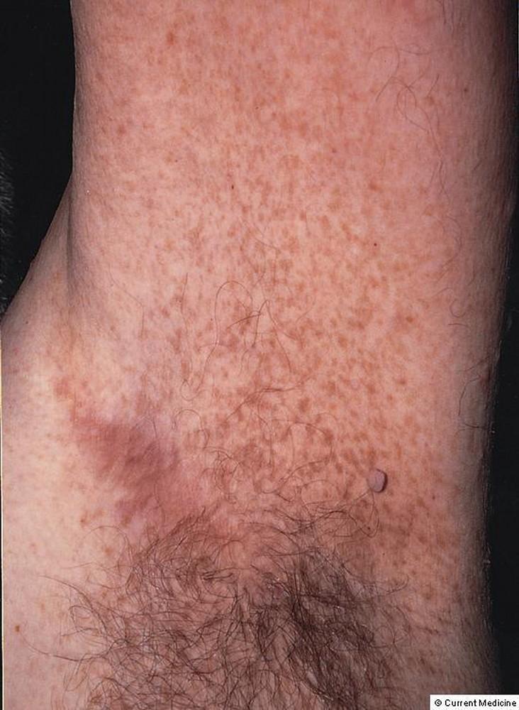
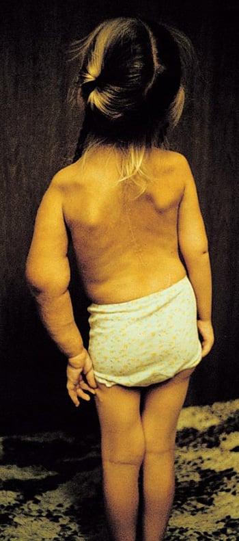
Café-au-lait lesions are medium-brown (café-au-lait), freckle-like macules, distributed most commonly over the trunk, pelvis, and flexor creases of elbows and knees. Although children who do not have neurofibromatosis may have 2 or 3 café-au-lait spots, children who have NF1 have ≥ 6 such macules and often many more. These macules are > 5 mm in affected prepubertal children and > 15 mm in postpubertal patients (see table Diagnosing Neurofibromatosis).
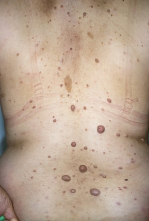
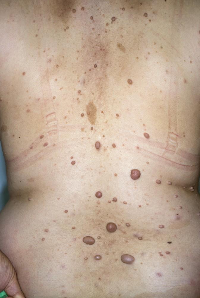
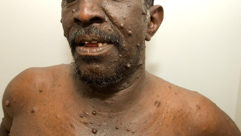
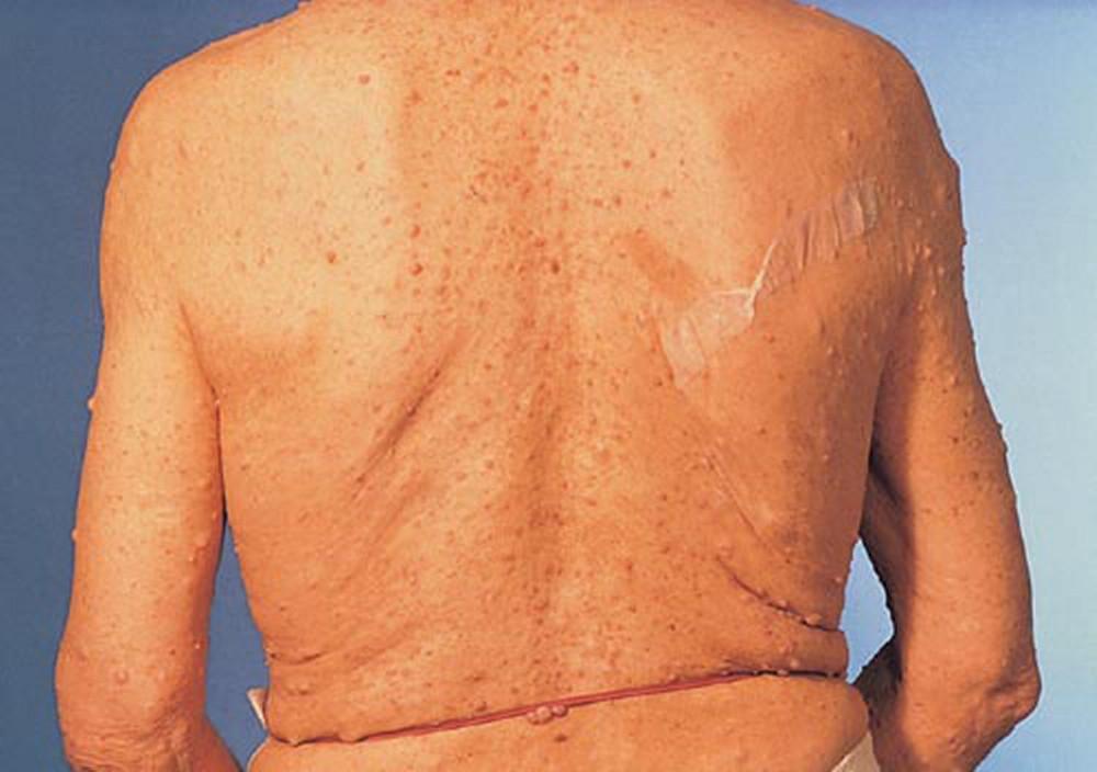
Cutaneous neurofibromas, which arise along small peripheral nerves, are common. During late childhood, these cutaneous tumors of various sizes and shapes appear, ranging in number from several to thousands. They may appear skin-colored or have a pink or tan discoloration and are usually asymptomatic.
Plexiform neurofibromas can develop and have a tendency to grow to large sizes, causing irregularly thickened, distorted structures, sometimes with grotesque deformities that can impinge on nerves and other structures. Plexiform neurofibromas can also involve cranial nerves, typically the 5th, 9th, and 10th.
Neurologic symptoms vary, depending on location and number of neurofibromas. Larger neurofibromas may press on their nerve of origin and cause distal paresthesia, pain, and sensory loss or weakness, depending on the function of the nerve that is involved. Neurofibromas that form along spinal nerve roots, especially where the nerve roots are contained by bone, can compress the nerve roots and cause radicular pain, weakness, or widespread sensory loss in that nerve distribution. Plexiform neurofibromas that compress cranial nerves cause deficits typical of those nerves.
DR HAROUT TANIELIAN/SCIENCE PHOTO LIBRARY

MEDICIMAGE / SCIENCE PHOTO LIBRARY

© Springer Science+Business Media

By permission of the publisher. From Bird T, Sumi S: Atlas of Clinical Neurology. Edited by RN Rosenberg. Philadelphia, Current Medicine, 2002.

DR HAROUT TANIELIAN/SCIENCE PHOTO LIBRARY

MEDICIMAGE / SCIENCE PHOTO LIBRARY

© Springer Science+Business Media

By permission of the publisher. From Bird T, Sumi S: Atlas of Clinical Neurology. Edited by RN Rosenberg. Philadelphia, Current Medicine, 2002.
Bone abnormalities include
Subperiosteal bone cysts
Vertebral scalloping
Thinning of the long-bone cortex
Pseudarthrosis
Absence of the greater wing of the sphenoid bone (posterior orbital wall), with consequent pulsating exophthalmos

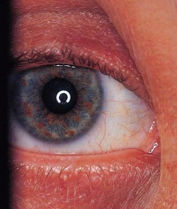
An optic glioma and Lisch nodules (iris hamartomas) occur in some patients. Optic gliomas are typically asymptomatic and do not require treatment unless they progressively increase in size.
Patients with NF1 can also have changes in their arterial walls that may lead to Moyamoya syndrome (stenosis or occlusion of arteries in and around the circle of Willis with formation of small collateral arteries) or intracranial aneurysms.
Some children have cognitive deficits, learning disorders, and slightly larger heads.

Children and adolescents with NF1 may have childhood chronic myelomonocytic leukemia (juvenile myelomonocytic leukemia) and rhabdomyosarcoma. Pheochromocytomas may occur at any age.
Malignant tumors are much less common but still more common than in the general population; they include supratentorial or brain stem gliomas and transformation of plexiform neurofibromas to malignant peripheral nerve sheath tumors. These tumors may develop at any age.
NF2-related schwannomatosis (NF2)
In NF2, bilateral vestibular schwannomas develop and become symptomatic during childhood or early adulthood. They cause hearing loss, unsteadiness, and sometimes headache or facial weakness. Bilateral 8th cranial (vestibulocochlear) nerve masses may be present.
Family members may have gliomas, meningiomas, or schwannomas.
Non-NF2 schwannomatosis (schwannomatosis)
In schwannomatosis, multiple schwannomas develop on cranial, spinal, and peripheral nerves. Vestibular schwannomas do not develop, and patients do not become deaf. Also, the other types of tumors that sometimes occur in neurocutaneous disorders do not develop.
The first symptom of schwannomatosis is usually pain, which may become chronic and severe. Other symptoms may develop, depending on the location of the schwannomas.
Diagnosis of Neurofibromatosis
Clinical evaluation
Brain MRI or head CT
Sometimes genetic testing
Most patients with NF1 are identified during routine examination, examination for cosmetic complaints, or evaluation of a positive family history.
Diagnosis of all 3 types is clinical (see table Diagnosing Neurofibromatosis) by detailed physical examination that is focused on the cutaneous, skeletal, and neurologic systems. NF1 should be suspected and monitored for in children who have multiple café-au-lait spots even if they do not have other features or family history of NF1.
Brain MRI is done in patients with neurologic symptoms or signs and, when detailed visual testing is not possible, in young children who meet the clinical criteria for NF1 and who may have an optic glioma. T2-weighted MRI may show thickening or tortuosity of the optic nerves and parenchymal hyperintense lesions that change over time and correlate with small cystic structures in NF1; MRI may help identify vestibular schwannomas or meningiomas in NF2. If vestibular schwannoma is suspected, CT of the petrous ridge can be done; it typically shows widening of the auditory canal.
Although the diagnosis can usually be established by clinical criteria, genetic testing is recommended for patients who are suspected to have neurofibromatosis but who do not fulfill clinical criteria.
Treatment of Neurofibromatosis
For symptomatic neurofibromas in NF1, possibly surgery or removal by laser or electrocautery
For malignant tumors, chemotherapy
For vestibular schwannomas, surgery and bevacizumab in selected cases
For non-NF2 schwannomatosis, primarily pain management
No general treatment for neurofibromatosis is available.
Most optic gliomas are asymptomatic and just need to be monitored for progression. For both progressive optic gliomas and central nervous system lesions that have become malignant, chemotherapy is the treatment of choice.
1). Hearing preservation and augmentation are equally important in the optimal care of these patients; hence all patients with NF2-related schwannomatosis should be referred to an audiologist.
Treatment of non-NF2 schwannomatosis is primarily symptomatic with long-term pain management. Surgical resection of schwannomas is recommended if the patient has uncontrolled pain or if the schwannomas cause neurologic deficit. Ideally, such patients are cared for by a multidisciplinary team with expertise in the various manifestations of the condition.
Genetic counseling is advisable for all types of neurofibromatosis. If either parent has neurofibromatosis, risk to subsequent children is 50%; if neither has it, risk for subsequent children is unclear because new mutations are common, particularly in NF1.
Treatment reference
1. Van Gompel JJ, Agazzi S, Carlson ML, et al: Congress of Neurological Surgeons Systematic Review and Evidence-Based Guidelines on Emerging Therapies for the Treatment of Patients With Vestibular Schwannomas. Neurosurgery 82(2):E52-E54, 2018. doi: 10.1093/neuros/nyx516
Key Points
There are 3 types of neurofibromatosis (NF): NF1, NF2-related schwannomatosis (previously NF2), and non-NF2 schwannomatosis (previously schwannomatosis), caused by gene mutations.
NF1 typically causes cutaneous, neurologic, and bone abnormalities but can affect almost any part of the body.
NF2 causes bilateral vestibular schwannomas.
Schwannomatosis causes multiple nonintradermal schwannomas; it does not cause vestibular schwannomas.
Diagnosis is made using clinical criteria; neuroimaging is done if patients have neurologic abnormalities.
Neurofibromas that cause severe symptoms may be removed surgically.
Malignant tumors may require chemotherapy.
Non-NF2 schwannomatosis requires symptomatic treatment with long-pain management.
Genetic testing is recommended for patients who are suspected to have neurofibromatosis but who do not fulfill clinical criteria.
More Information
The following English-language resources may be useful. Please note that THE MANUAL is not responsible for the content of these resources.
United Kingdom Neurofibromatosis Association Clinical Advisory Board: Guidelines for the diagnosis and management of individuals with NF1 (2007)
Guidelines for the health supervision of children with NF1 from a collaboration of experts (2019)
American College of Medical Genetics and Genomics: Clinical practice guidelines for the care of adults with NF1 (2018)


