


Gas exchange is measured through several means, including
Diffusing capacity for carbon monoxide
Pulse oximetry
Arterial blood gas sampling
Diffusing Capacity for Carbon Monoxide
The diffusing capacity for carbon monoxide (DLCO) is a measure of the ability of gas to transfer from the alveoli across the alveolar epithelium and the capillary endothelium to the red blood cells. The DLCO depends not only on the area and thickness of the blood-gas barrier but also on the volume of blood in the pulmonary capillaries. The distribution of alveolar volume and ventilation also affects the measurement.
DLCO is measured by sampling end-expiratory gas for carbon monoxide (CO) after patients inspire a small amount of carbon monoxide, hold their breath, and exhale. Measured DLCO should be adjusted for alveolar volume (which is estimated from dilution of helium) and the patient’s hematocrit. DLCO is reported as mL/minute/mm Hg and as a percentage of a predicted value.
Pulse Oximetry
Transcutaneous pulse oximetry estimates oxygen saturation (SpO2) of capillary blood based on the absorption of light from light-emitting diodes positioned in a finger clip or adhesive strip probe. The estimates are generally very accurate and correlate to within 5% of measured arterial oxygen saturation (SaO2). Results may be less accurate in patients with
Highly pigmented skin
Arrhythmias
Hypotension
Profound systemic vasoconstriction
Pulse oximetry results are also less accurate in patients wearing nail polish.
Pulse oximetry is able to detect only oxyhemoglobin or reduced hemoglobin but not other types of hemoglobin (eg, carboxyhemoglobin, methemoglobin); those types are assumed to be oxyhemoglobin and falsely elevate the SpO2 measurement.
Arterial Blood Gas (ABG) Sampling
ABG sampling is done to obtain accurate measures of arterial oxygen partial pressure (PaO2), arterial carbon dioxide partial pressure (PaCO2), and arterial pH; these variables adjusted for the patient’s temperature allow for calculation of bicarbonate level (which can also be measured directly from venous blood) and SaO2. ABG sampling can also accurately measure carboxyhemoglobin and methemoglobin.
The radial artery is usually used. Because arterial puncture in rare cases leads to thrombosis and impaired perfusion of distal tissue, Allen test may be done to assess adequacy of collateral circulation. With this maneuver, the radial and ulnar pulses are simultaneously occluded until the patient's hand becomes pale. The ulnar pulse is then released while pressure on the radial pulse is maintained. A blush across the entire hand within 7 seconds of release of the ulnar pulse suggests adequate flow through the ulnar artery.
Under sterile conditions, a 22- to 25-gauge needle attached to a heparin-treated syringe is inserted just proximal to the maximal impulse of the radial arterial pulse and advanced slightly distally into the artery until pulsatile blood is returned. Systolic blood pressure is usually sufficient to push back the syringe plunger. After 3 to 5 mL of blood is collected, the needle is quickly withdrawn, and firm pressure is applied to the puncture site to facilitate hemostasis. Simultaneously, the ABG specimen is placed on ice to reduce oxygen consumption and carbon dioxide production by WBCs and is sent to the laboratory.
Oxygenation
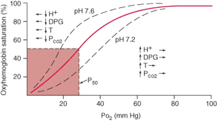
Hypoxemia is a decrease in the partial pressure of oxygen (PO2) in arterial blood; hypoxia is a decrease in the PO2 in the tissue. ABGs accurately assess the presence of hypoxemia, which is generally defined as a PaO2 low enough to reduce the SaO2 below 90% (ie, PaO2 < 60 mm Hg). Abnormalities in hemoglobin (eg, methemoglobin), higher temperatures, lower pH, and higher levels of 2,3-diphosphoglycerate reduce hemoglobin SaO2 despite an adequate PaO2, as indicated by the oxyhemoglobin dissociation curve.
Oxyhemoglobin dissociation curve
Arterial oxyhemoglobin saturation is related to PO2. PO2 at 50% saturation (P50) is normally 27 mm Hg. The dissociation curve is shifted to the right by increased hydrogen ion (H+) concentration, increased red blood cell 2,3-diphosphoglycerate (DPG), increased temperature (T), and increased PCO2. Decreased levels of H+, DPG, temperature, and PCO2 shift the curve to the left. Hemoglobin characterized by a rightward shifting of the curve has a decreased affinity for oxygen, and Hemoglobin characterized by a leftward shifting of the curve has an increased affinity for oxygen. |

Causes of hypoxemia can be classified based on whether the alveolar-arterial PO2 gradient [(A-a)DO2], defined as the difference between alveolar oxygen tension (PAO2) and PaO2, is elevated or normal. PAO2 is calculated as follows:

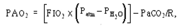
where FIO2 is the fraction of inspired oxygen (eg, 0.21 at room air), Patm is the ambient barometric pressure (eg, 760 mm Hg at sea level), PH2O is the partial pressure of water vapor (eg, usually 47 mm Hg), PaCO2 is the measured partial pressure of arterial carbon dioxide, and R is the respiratory quotient, which is assumed to be 0.8 in a resting patient eating a normal diet.
For patients at sea level and breathing room air, FIO2 = 0.21, and the (A-a)DO2 can be simplified as follows:


where (A-a)DO2 is typically < 20 but increases with age (because of age-related decline in pulmonary function) and with increasing FIO2 (because, although hemoglobin becomes 100% saturated at a PaO2 of about 150 mm Hg, oxygen is soluble in blood, and the oxygen content of plasma continues to increase at increasing FIO2). Estimations of normal (A-a)DO2 values as < (2.5 + [FIO2× age in years]) or as less than the absolute value of the FIO2 (eg, < 21 while breathing room air; < 30 on 30% FIO2) correct for these effects.
Hypoxemia with increased (A-a)DO2
Hypoxemia with increased (A-a)DO2 is caused by
Low ventilation/perfusion (V/Q) ratio (a type of V/Q mismatch)
Right-to-left shunting
Severely impaired diffusing capacity
Low V/Q ratio is one of the more common reasons for hypoxemia and contributes to the hypoxemia occurring in COPD and asthma. In normal lungs, regional perfusion closely matches regional ventilation because arteriolar vasoconstriction occurs in response to alveolar hypoxia. In disease states, dysregulation leads to perfusion of alveolar units that are receiving less than complete ventilation (V/Q mismatch). As a result, systemic venous blood passes through the pulmonary capillaries without achieving normal levels of PaO2. V/Q mismatch can also occur when there is increased blood flow even when ventilation is normal, as in liver disease. Supplemental oxygen can correct hypoxemia due to low V/Q ratio by increasing the PAO2, although the increased (A-a)DO2 persists.
Right-to-left shunting is an extreme example of low V/Q ratio. With shunting, deoxygenated pulmonary arterial blood arrives at the left side of the heart without having passed through ventilated lung segments. Shunting may occur through lung parenchyma, through abnormal connections between the pulmonary arterial and venous circulations, or through intracardiac communications (eg, patent foramen ovale). Hypoxemia due to right-to-left shunting does not respond to supplemental oxygen.
Impaired diffusing capacity only rarely occurs in isolation; usually it is accompanied by low V/Q ratio. Because oxygen completely saturates hemoglobin after only a fraction of the time that blood is in contact with alveolar gas, hypoxemia due to impaired diffusing capacity occurs only when cardiac output is increased (eg, during exercise), when barometric pressure is low (eg, at high altitudes), or when > 50% of the pulmonary parenchyma is destroyed. As with low V/Q ratio, the (A-a)DO2 is increased, but PaO2 can be increased by increasing the FIO2. Hypoxemia due to impaired diffusing capacity responds to supplemental oxygen.
Hypoxemia with normal (A-a)DO2
Hypoxemia with normal (A-a)DO2 is caused by
Hypoventilation
Low partial pressures of inspired oxygen (PIO2)
Hypoventilation (reduced alveolar ventilation) decreases the PAO2 and increases the PaCO2, thereby decreasing PaO2. In cases of pure hypoventilation, the (A-a)DO2 is normal. Causes of hypoventilation include decreased respiratory rate or depth (eg, due to neuromuscular disorders, severe obesity, or drug overdose, or in compensation for metabolic alkalosis) or an increase in the fraction of dead space ventilation in patients already at their maximal ventilatory limit (eg, an exacerbation of severe COPD). Hypoventilatory hypoxemia responds to supplemental oxygen.
Decreased PIO2 is an uncommon cause of hypoxemia that in most cases occurs only at high altitude. Although FIO2 does not change with altitude, ambient air pressure decreases exponentially; thus, PIO2 decreases as well. For example, PIO2 is only 43 mm Hg at the summit of Mt. Everest (altitude, 8848 m [29,028 ft]). The (A-a)DO2 remains normal. Hypoxic stimulation of respiratory drive increases alveolar ventilation and decreases PaCO2 level. This type of hypoxemia responds to supplemental oxygen.
Carbon Dioxide
PCO2 normally is maintained between 35 and 45 mm Hg. A dissociation curve similar to that for oxygen exists for carbon dioxide but is nearly linear over the physiologic range of PaCO2. Abnormal PCO2 is almost always linked to disorders of ventilation (unless occurring in compensation for a metabolic abnormality) and is always associated with acid-base changes.
Hypercapnia
Hypercapnia is PCO2 > 45 mm Hg. Causes of hypercapnia are the same as those of hypoventilation (eg, disorders that decrease respiratory rate or depth or increase the fraction of dead space ventilation in patients already at their maximal ventilatory limit). Disorders that increase carbon dioxide production (eg, hyperthyroidism, fever) when combined with an inability to increase ventilation also cause hypercapnia.
Hypocapnia
Hypocapnia is PCO2 < 35 mm Hg. Hypocapnia is always caused by hyperventilation due to pulmonary (eg, pulmonary edema, pulmonary embolism), cardiac (eg, heart failure), metabolic (eg, acidosis
Carboxyhemoglobinemia
Carbon monoxide binds to hemoglobin with an affinity 210 times that of oxygen and prevents oxygen transport. Clinically toxic carboxyhemoglobin levels are most often the result of exposure to exhaust fumes or from smoke inhalation, although cigarette smokers have detectable levels.
Patients with carbon monoxide poisoning may present with nonspecific symptoms such as malaise, headache, and nausea. Because poisoning often occurs during colder months (because of indoor use of combustible fuel heaters), symptoms may be confused with a viral syndrome such as influenza. Clinicians must be alert to the possibility of carbon monoxide poisoning and measure levels of carboxyhemoglobin when indicated. Carboxyhemoglobin can be directly measured from venous blood—an arterial sample is unnecessary. Oxygen saturation determined by pulse oximetry will be normal and cannot be used to screen for carbon monoxide poisoning. Carboxyhemoglobin can be measured by co-oximetry.
Treatment is the administration of 100% oxygen (which shortens the half-life of carboxyhemoglobin) and sometimes the use of a hyperbaric chamber.
Pearls & Pitfalls
|
Methemoglobinemia
Methemoglobin is hemoglobin in which the iron is oxidized from its ferrous (Fe2+) to its ferric (Fe3+) state. Methemoglobin does not carry oxygen and shifts the normal oxyhemoglobin dissociation curve to the left, thereby limiting the release of oxygen to the tissues.
Methemoglobin level can be directly measured by co-oximetry (which emits 4 wavelengths of light and is capable of detecting methemoglobin, carboxyhemoglobin, hemoglobin, and oxyhemoglobin) or may be estimated by the difference between the oxygen saturation calculated from the measured PaO2 and the directly measured oxygen saturation. Oxygen saturation measured by pulse oximetry will be inaccurate in the presence of methemoglobinemia.


