


Coccidiosis is a worldwide ovine problem caused by the genus Eimeria, protozoal intracellular parasites of the intestinal epithelial cells. They cause considerable disease and economic loss. Under most sheep production systems, all, or nearly all, animals are exposed to some Eimeria spp, although most infections are inapparent.
Etiology of Coccidiosis of Sheep
At least 13 species of Eimeria infect sheep: Eimeria ahsata, E bakuensis, E crandallis,E faurei, E gilruthi (previously Globidium gilruthi), E granulosa, E intricata, E marsica, E ovina (E arloingi A), E ovinoidalis (E ninokohlyakimovae),E pallida, E parva, and E weybridgensis (E arloingi B). However, only 2 are considered to be pathogenic—E crandallis and E ovinoidalis (most pathogenic). Except for E gilruthi, all sheep coccidial species are considered host-specific (stenoxenous development) and so do not also infect cattle, goats, or poultry. Often concurrent mixed Eimeria species are present in the host and may increase the severity of any signs. There is no cross-immunity between different ovine Eimeria species. Because the most susceptible age to infection is lambs, the helminth Nematodirus battus may also contribute to any clinical signs.
The complicated life cycle of sheep Eimeria species takes 2 to 3 weeks to complete (prepatent period). After fecal shed, oocysts are not infective until they sporulate, which requires several days in moderately warm and moist conditions. Oocysts are relatively resistant to disinfectants but are killed by desiccation or temperatures >55º–60ºC. Infective oocysts can overwinter. The life cycle of E gilruthi is uncertain.
Almost all Eimeria species infect cells in preferential sites in the small or large intestine. For example, E crandallis infects the ileum, but E ovinoidalis may also infect the cecum and colon, possibly resulting in bloody diarrhea. The lower intestinal areas are concerned with water and feed absorption, and, as cell replacement is slow, there are no compensatory effects during infection. In such cases, there is decreased water absorption and severe hemorrhage where the large intestinal mucosa is denuded. This can lead to dehydration from decreased fluid retention and can cause decreased circulating phosphorus, potassium, and sodium concentrations. Those that do not die often have long recovery periods and may remain stunted or with decreased growth rates. Infection with E gilruthi occurs in the abomasum but much of its pathogenesis is unknown.
Epidemiology of Coccidiosis of Sheep
When sheep are born outside under extensive or nomadic conditions, clinical illness is rare. However, when lambing outside involves lambing pens or small areas, infection can build up. Usually, lambs born indoors are all quickly exposed to infection and so they are more vulnerable to clinical disease. It seems probable that most lambs are infected by ewes or older sheep. There is argument as to whether or not ewes show a periparturient oocyst increase with shedding towards lambing, although oocyst counts tend to rise as lambing approaches. Fecal contamination of the environment allows oocysts to be present in lambing pens (especially when uncleaned or partially cleaned), bedding, and feed. The udders and teats of ewes may become soiled, plus small numbers in the ewe’s feces allow early infection exposure of the lamb. Infections in the adult are usually without clinical signs. Although sheep of any age can be affected, most clinical cases occur between 1 and 6 months of age, with many occurring between 1 and 2 months of age in indoor lambs.
In North America, disease often occurs at the time of transition from winter to spring. Intensive grazing areas and feedlots are at greatest risk as a result of shipping, ration change, crowding stress, severe weather, and contamination of the environment with oocysts from ewes or other lambs.
After exposure, immunity is good and is enhanced by continual exposure to low levels of infection. Under natural conditions, lambs pick up a small continuous infection and become immune without any clinical signs. Small numbers of oocysts are shed throughout the sheep’s life, and numbers increase towards parturition.
Clinical Signs of Coccidiosis of Sheep
Most lambs become infected but show no clinical disease. Most clinical disease occurs in lambs 4–8 weeks old reared in a contaminated environment (indoors or outside). When lambs are all of a similar age, most may show clinical signs. Acute infections result in sudden onset diarrhea, anorexia, dullness, and abdominal pain. Dehydration may result and there is marked loss of weight and body condition. Fever is unusual. Severe acute infections may lead to diarrhea before any oocyst shedding and, especially with E ovinoidalis, it may be bloody. Chronic infections result in a reduction in feed consumption, feed conversion, and growth rates.

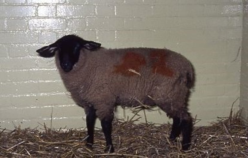
Courtesy of Dr. Anthony Andrews.

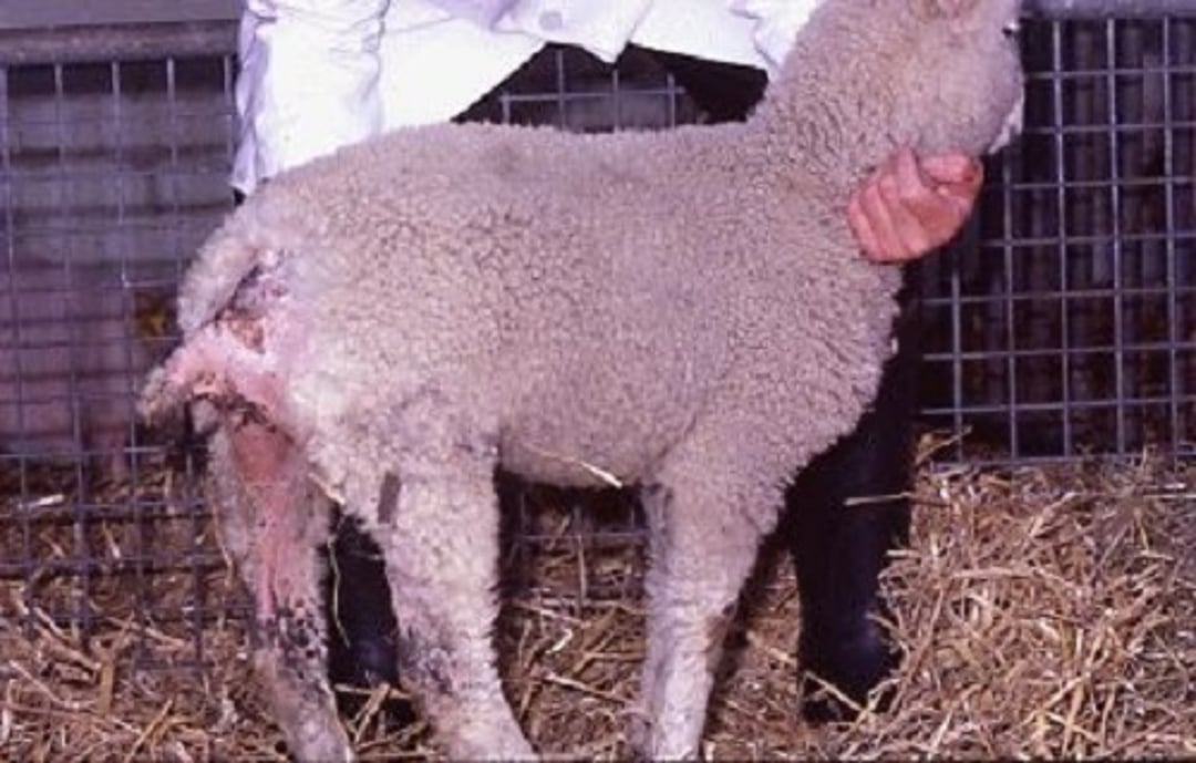
Courtesy of Anthony Andrews.
Postmortem Lesions
Depending on the Eimeria species, infection is in the small or large intestine. The ileum, cecum, and upper colon are usually most affected and may be thickened, edematous, and inflamed; sometimes, there is mucosal hemorrhage. Of the two main pathogenic species, E crandallis and E ovinoidalis have life cycles involving large numbers of cells becoming parasitized during the developmental stages, resulting in the intestinal epithelium being gradually eroded; in some cases there is widespread damage. This can lead to secondary bacterial infection. E ahsata causes catarrhal enteritis; E bakuensis produces mucosal polyps formation; E crandallis results in localized epithelial shedding; E faurei also causes catarhal enteritis; E ovina can cause raised visible white, opaque patches containing large numbers oocysts in the small intestine; E ovinoidalis lesions are hemorrhagic colitis and typhlitis. Histologic examination shows loss of intestinal epithelial cells, with villous atrophy and crypt hyperplasia. E gilruthi produces an abomasitis, with edema, hemorrhages, and raised nodules of the abomasal mucosa. Histologically, the nodules comprised the organisms surrounded by an area of necrosis.
Diagnosis of Coccidiosis of Sheep
Diagnosis is based on several factors, including age, history, presence of oocysts in feces, oocyst speciation, and any postmortem findings. Severe diarrhea in 4–8 week old lambs indoors or on highly stocked pastures is an indication. Fecal samples should be taken from a sample of lambs with and without diarrhea.
A fecal oocyte count >5,000 oocysts/g with appropriate signs may be noteworthy, but speciation should be undertaken. The feces usually contains many oocysts, which can be detected by flotation techniques (saturated salt or glucose solution). Oocysts and N battus eggs may both be present and indicate more severe infections. Coccidial oocyst count can be complicated by animal selection, timing of sampling, presence of nonpathogenic Eimeria spp and lack of agreement on interpretation of noteworthy oocyst counts. Speciation should be undertaken with 2% potassium dichromate as E crandallis and E ovinoidalis are of similar size. Postmortem findings show intestinal damage with white meront patches. Smears show various development stages including meronts, oocysts, and others. Histologic findings also indicate parasite presence.
If clinical signs are in lambs from about a month onward, most problems can be differentiated by examination of fecal samples selected from animals with and without clinical signs. Salmonella spp or other bacteria can be determined by bacteriologic culture. Clostridiumperfringens type C and D can be detected on smears and possible toxin identification. Coronavirus and rotavirus can be shown by ELISA. Cryptosporidia can be detected by oocyst examination or ELISA. Helminths are indicated by presence of eggs (Nematodirus battus occurs in lambs this age and has relatively large eggs). Otherwise, digestive, nutritional, and management problems can usually be determined via careful history-taking and observation, feed examination, and possibly blood tests.
Treatment of Coccidiosis of Sheep
All severely infected lambs should be separated from their mothers and placed in a clean, disinfected pen with abundant straw. Those affected outside should be moved to less contaminated, well-drained areas. Ideally, the remaining in-contact sheep inside or outdoors are also moved to less contaminated areas to ensure feed and water sources can be kept free of feces. Depending on the circumstances, it may be necessary to treat all lambs without clinical signs. In the US, few treatments are approved for use in sheep and most are prescribed for extralabel usage. Medicines are only effective if treatment begins early in the outbreak. Clinically affected animals should receive individual treatment. Those severely affected may need electrolyte and nutritional supplementation.
Sulfaquinoxaline in drinking water (0.015% concentration for 3–5 days) may be used to treat affected lambs. Sulfamethazine (sulfadimidine) can be administered at 247.5 mg/kg the first day, then 124 mg/kg for 3 days. Sulfadimethoxine is administered at 55 mg/kg the first day, then 27.5 mg/kg for 3 days, by mouth or in the drinking water. All three medicines are approved for some ruminant species but not sheep. Because water intake is often decreased, ill animals should be medicated by drenching.
Diclazuril (1 mg/kg, PO, once, or repeated 3 weeks later) is licensed in some countries. Toltrazuril (20 mg/kg, PO, once) is used to decrease oocyst counts and is licensed in some countries. Ponazuril, a metabolite of toltrazuril, is suggested for control in sheep and used experimentally in goat kids. Currently, no product is licensed for food animals. Currently, ponazuril is indicated for equine protozoal myeloencephalitis.
Decoquinate (1 mg/kg, PO in feed, every 24 hours for 28 days) is licensed in some countries, including the US, for young nonlactating sheep; using a 60 g/kg premix at 1.67 kg/ton of feed provides the recommended 100 mg/kg of feed, allowing 100 g of feed for a 10-kg lamb for 28 days. However, if lamb feed intake is less, higher premix concentrations are necessary. Decoquinate may prevent decreased neutrophil function due to the parasite.
Amprolium (50–55 mg/kg, PO, every 24 hours for 5 days) can be used in an extralabel fashion in the US and some other countries and should be under veterinary control. Use in lambs is controversial as it can cause thiamine deficiency and associated neurologic disease. As a precaution, thiamine (vitamin B1) is also often provided after treatment.
Control and Prevention of Coccidiosis of Sheep
Management can assist in preventing disease by decreasing stocking density, decreasing fecal contamination from ewes or lambs, and decreasing potential stresses, such as those from shipping, ration change, and severe weather. All feed and water troughs should be raised from the ground and fecal contamination prevented. All troughs inside and outside should be sited in well-drained areas. Regular movement of lambs to new areas prevents the excessive buildup of oocysts. Lambs should be weaned when the weather is stable and kept on a consistent diet. Desiccation and high temperatures (>55º–60ºC) kill off the oocysts.
Some ammonia-based disinfectants destroy the oocysts but cannot be used while animals are present. Good nutrition for the pregnant ewe produces strong healthy lambs and provides sufficient high quality colostrum, thereby decreasing coccidial problems.
Occurrence of coccidiosis under many management systems is predictable, so in-feed or soluble coccidiostats may need to be administered prophylactically for 28 consecutive days, beginning a few days after lambs are introduced into the suspect environment. However, this does not mean that other suitable remedial management changes should not be sought and introduced. Sulfonamides can be added to the drinking water as for treatment but are not approved by the FDA for this purpose. Their inclusion may decrease or stop drinking.
A combination of monensin and lasalocid at 22 and 100 mg/kg of diet, respectively, may be an effective prophylactic against naturally occurring coccidiosis in early-weaned lambs under feedlot conditions.
Amprolium (15 mg/kg, PO, every 24 hours for 19–21 days) is used as an extralabel preventive in the US during periods of exposure or when conditions are likely to result in problems.
Monensin is used in the US as concentrated rations at 15 g/ton and is fed to ewes from 4 weeks before lambing until weaning, and to lambs from 4–20 weeks of age. The toxic level for lambs is 4 mg/kg but it is only FDA-approved for confined goats. Lasalocid (15–70 mg per sheep per day, depending on body wt) may be effective and is FDA-approved for confined sheep. A combination of monensin and lasalocid at 22 and 100 mg/kg of diet, respectively, may be an effective prophylactic against naturally occurring coccidiosis in early weaned lambs under feedlot conditions.
Decoquinate in feed is approved in many countries for coccidial control in lambs and is fed at the same dosage as for treatment (1 mg/kg, PO in feed, every 24 hours for 28 days). It is also effective in decreasing oocyst shedding when fed continually to ewes in dry feed at 0.5 mg/kg (using a 60 g/kg premix at 833 g/ton of feed provides the recommended treatment dose of 50 mg/kg of feed). It should be provided for at least 28 days. Inclusion rates may need to increase if feed intake is insufficient.
Toltrazuril (20 mg/kg, PO, once) is licensed in many countries for use before anticipated oocyst shedding. Diclazuril (1 mg/kg, PO, once, with a possible second dose 3 weeks later) is also licensed in some countries for sheep 4–6 weeks of age; it can control infection. Ponazuril is suggested to decrease oocyst counts, but the product available is licensed in the US for protozoal myeloencephalitis in horses.
Natural products have been recorded as having some beneficial effects. Oregano oil may aid in coccidiosis control. Lespedeza cuneata in feed pellets have decreased fecal signs and oocyst counts in goat kids.
There are no vaccines available.


