


Esophageal foreign bodies are more common in dogs than in cats. Bones are the most common foreign body; however, needles, fishhooks, wood, rawhide, and dental chew treats may also become lodged in the esophagus. Objects usually lodge in the areas of the esophagus with the least distensibility: the thoracic inlet, over the heart base, or the caudal esophagus just cranial to the diaphragm. Occasionally, an object may lodge in other locations such as the upper esophageal sphincter.
Ptyalism, gagging, dysphagia, regurgitation, and repeated attempts to swallow are clinical signs of an esophageal foreign body. Often, the owner may see the animal eat the foreign body. The clinical signs depend on the location of the foreign body and on the extent and duration of obstruction. A partial obstruction may allow fluids but not food to pass. With a chronic obstruction, anorexia, weight loss, and lethargy are common.
Perforation of the cervical esophagus may result in local abscessation or subcutaneous emphysema; perforation of the thoracic esophagus may result in pleuritis, mediastinitis, pyothorax, pneumothorax, bronchoesophageal fistula formation, or fatal aortic esophageal fistula formation. Esophagitis, mucosal laceration, esophageal stricture, and esophageal diverticulum formation are also potential complications. Esophageal stricture formation is the most common complication associated with an esophageal foreign body. Aspiration pneumonia may also be present secondary to the regurgitation.
Many esophageal foreign bodies are radiopaque and can be seen on plain radiographs. A contrast esophagram or esophagoscopy is often required to identify radiolucent foreign bodies. Because barium paste can obscure and potentially damage an endoscope, waiting 24 hours after an esophagram is recommended before doing an esophagoscopy. If a perforation is suspected, an iodinated contrast medium should be used instead of barium suspensions. Esophagoscopy permits evaluation of both the foreign body and the esophageal wall and often allows therapeutic intervention.

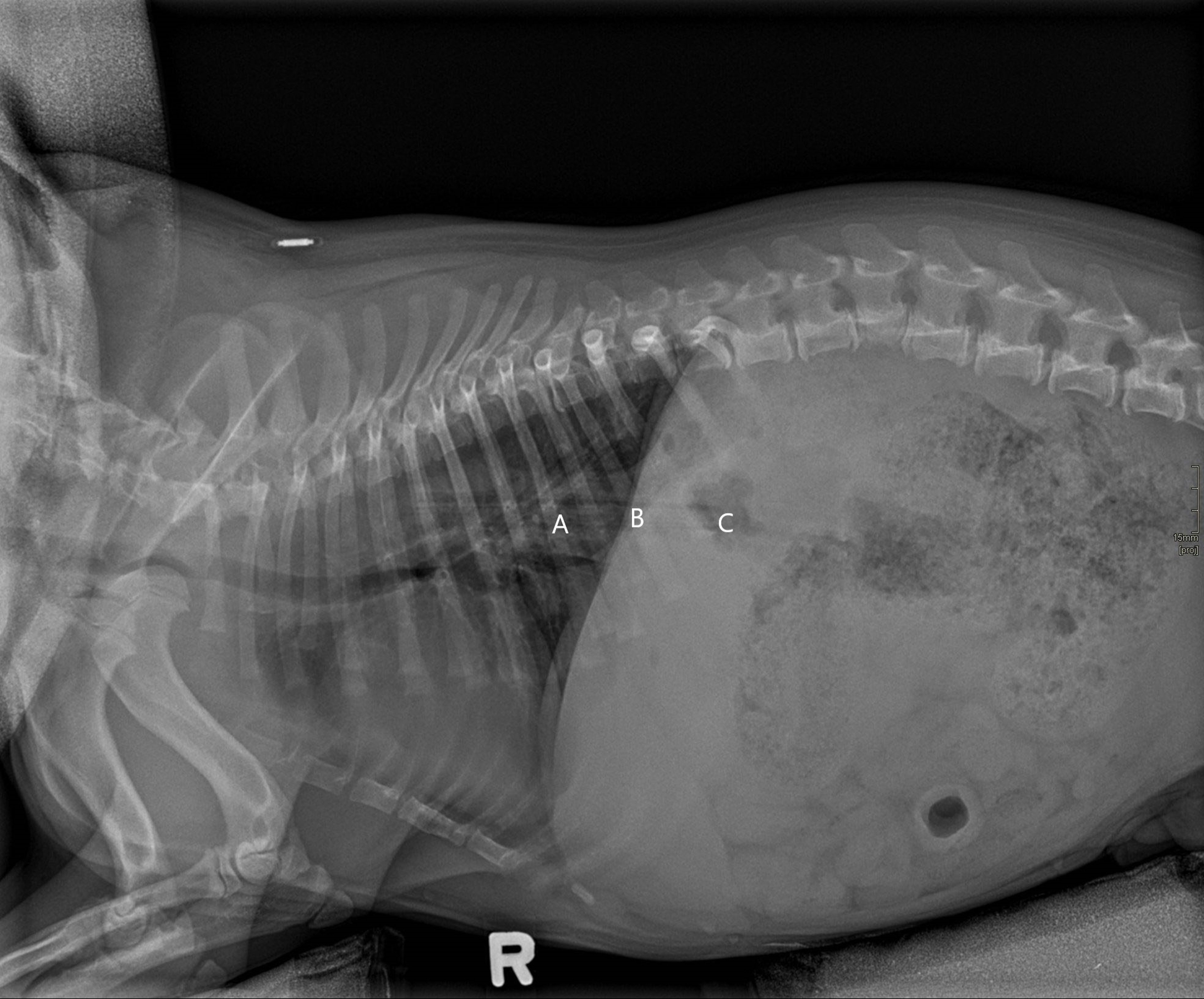
Courtesy of Alex Zur Linden.
Esophageal foreign bodies, once diagnosed, should be removed immediately. Most often, a foreign body can be removed per os with a flexible endoscope and forceps. A rigid endoscope can also be used if a flexible scope is not available; however, care must be taken when manipulating the scope in the esophagus to prevent lacerations or perforations. If the foreign body is smooth, a Foley catheter can be inserted distal to the foreign body, inflated, then removed orally, bringing the foreign body with it. A large endotracheal tube can be placed over the endoscope to remove sharp foreign bodies such as fish hooks, which can be drawn up into the endotracheal tube and removed without damaging the esophagus on the way out.
If a foreign body cannot be removed per os, it may be pushed into the stomach, where it can either be digested (eg, bones), passed, or removed via a gastrotomy. Surgery is indicated if a perforation has occurred or the foreign body cannot be removed via endoscopy. However, there is potential for stricture formation and complications secondary to the poor wound-healing ability of the esophagus. Esophagitis, if present, should be treated appropriately.
If severe circumferential damage of the esophagus is observed, the risk of stricture formation is higher, and a recheck endoscopy should be recommended within 1–2 weeks after foreign body removal. Percutaneous endoscopic gastrotomy tube placement while removing the foreign body is controversial. Keeping the esophagus opened mechanically via oral feeding may be beneficial. However, feeding may trigger more inflammation, damage, and pain. In all cases, aggressive medical management, including administering sucralfate and omeprazole, is recommended.


