


The condition commonly referred to as "gastric ulcers" is pigs more accurately described as gastroesophageal ulceration. Lesions affect the esophagus and the cranial, nonglandular region of the stomach (pars esophagea). Lesions can vary from an initial thickening (hyperkeratosis) of the surface of the pars esophagea, also known as parakeratosis which progresses to fissures and gastric ulceration. Lesions may be localized or can involve the entire surface of the pars esophagea. Clinical signs are related to blood loss associated with ulceration. Sudden death may occur. In such cases, typically, the carcass is pale but otherwise in good body condition. Treatment of surviving pen-mates generally involves changes in feed, specifically the introduction of coarsely-ground feed (ie, large particle size); alteration of housing and management to ensure feed intake is not interrupted or limited; or a combination of these interventions.
Etiology of Gastric Ulcers in Pigs
Ulcers may affect the pars esophagea in pigs and cause sporadic cases of acute gastric hemorrhage and death or slow growth due to chronic ulceration. Ulcers result when the unprotected stratified squamous epithelium of the pars esophagea is subjected to insult from the mixture of acid, bile, and digestive enzymes present in the distal region of the stomach. The mucosa that surrounds the esophageal opening is not protected by mucus and therefore relies on maintenance of a pH gradient between the proximal and distal regions of the stomach in pigs.
Many risk factors are associated with the development of ulcers, most of which are associated with an increase in fluidity of the stomach contents. The most important of these factors relate to feed, particularly the use of finely ground feed (< 700 microns average particle size). Consumption of finely ground pelleted feed results in rapid stomach transit time and fluid stomach contents. Other important factors tend to be associated with disruption in feed intake, eg, an outbreak of respiratory disease, hot weather, or management errors that lead to empty feeders. Disruption in feed intake also results in fluid stomach contents and lack of a physical barrier between the acidic distal contents and the sensitive pars esophageal region.
Factors that cause increased acid production may possibly influence the prevalence and severity of ulcers, but there is little evidence that hyperacidity is a primary cause. Similarly, Helicobacter-like organisms can often be identified under the mucus on the surface of the glandular mucosa of the distal stomach, but evidence that these bacteria are a cause of ulcer development in the pars esophagea is lacking, and epidemiologic studies showing an association are inconsistent. The presence of the bacteria in the glandular region and the lesion in the pars esophagea is difficult to explain from a biological standpoint.
Clinical Findings of Gastric Ulcers in Pigs

Courtesy of Dr. Robert Friendship.
Courtesy of Dr. Robert Friendship.
Courtesy of Dr. Robert Friendship.
Courtesy of Dr. Robert Friendship.
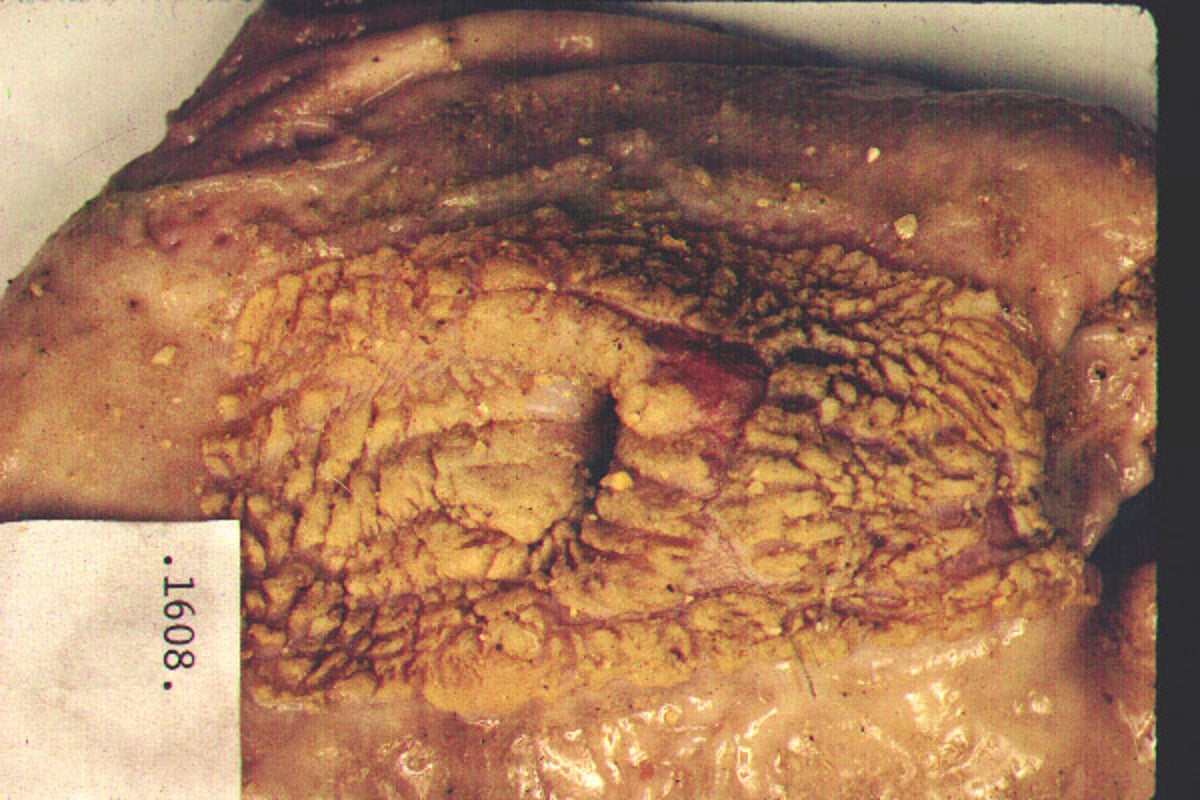
Sudden death usually involving only a small number of pigs in a grower-finisher barn is a common presentation of gastric ulcers, but higher numbers can occur sporadically and are usually associated with a triggering event such as feed disruption. Death is caused by acute hemorrhage from the ulcer into the stomach, so the carcass appears notably pale but generally in good body condition. If bleeding is less severe, the clinical signs are those associated with anemia, such as pallor. If bleeding is prolonged, affected pigs become weak, lose body condition, and grow more slowly than pen-mates. Scant amounts of black tarry feces indicating digested blood may be noted. Vomiting may be seen but diarrhea is generally not present, which helps to differentiate gastric ulceration from other conditions. Other causes of blood loss or anemia need to be excluded.
The most common age group to show clinical signs are pigs 2–6 months old, as well as sows. Most pigs reared in modern confinement systems develop lesions in the pars esophagea. Lesions can both develop and heal quickly. This makes it difficult to determine whether erosions of the pars esophageal mucosa cause delayed growth if blood loss is minimal and anemia is not present. Scarring can occur and, if severe, can result in narrowing of the gastroesophageal junction. Clinical signs of esophageal stenosis include regurgitation of feed shortly after consumption and loss of body condition.
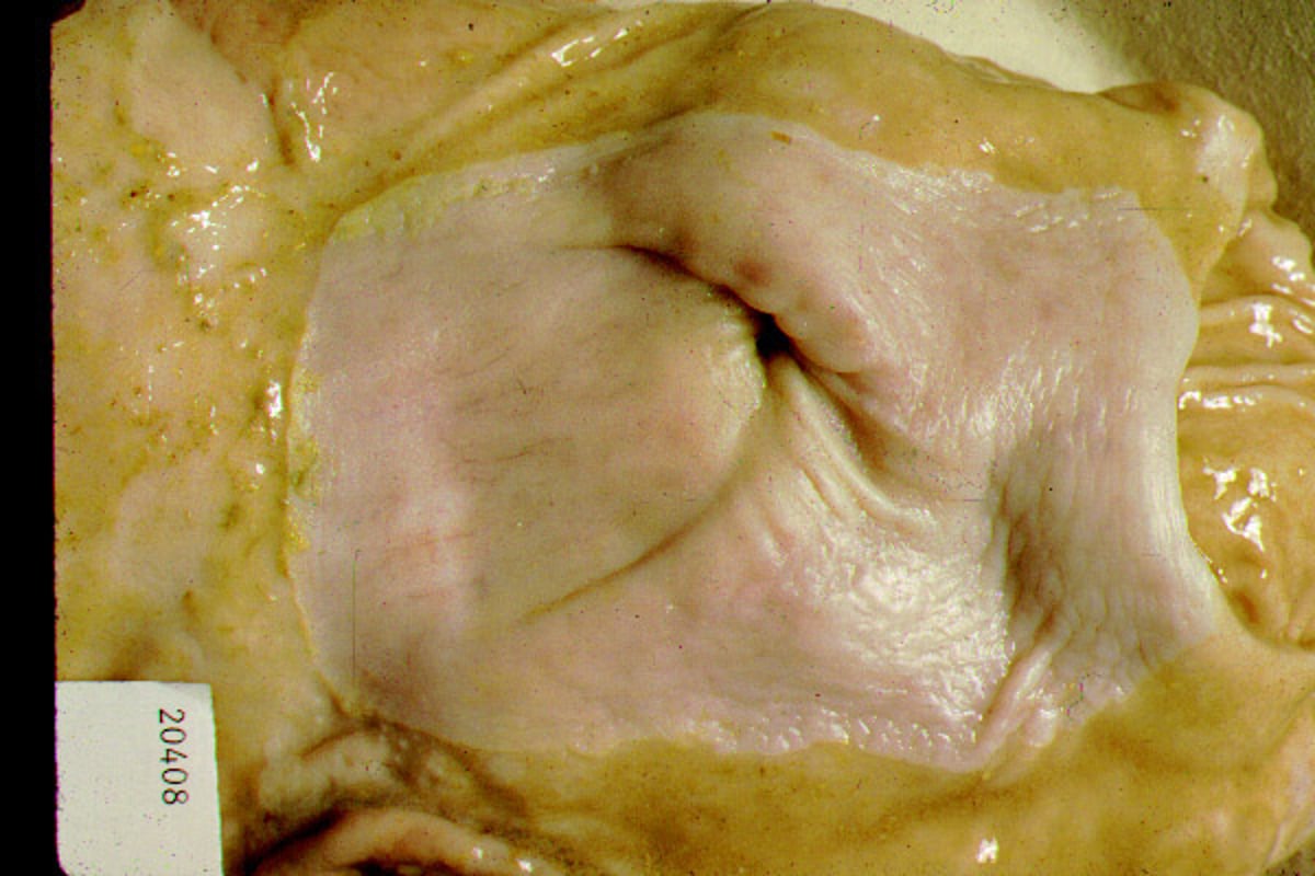
Lesions
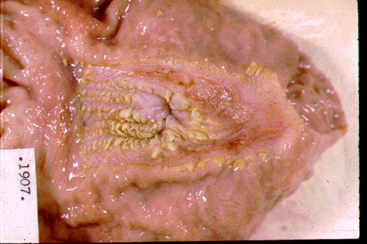
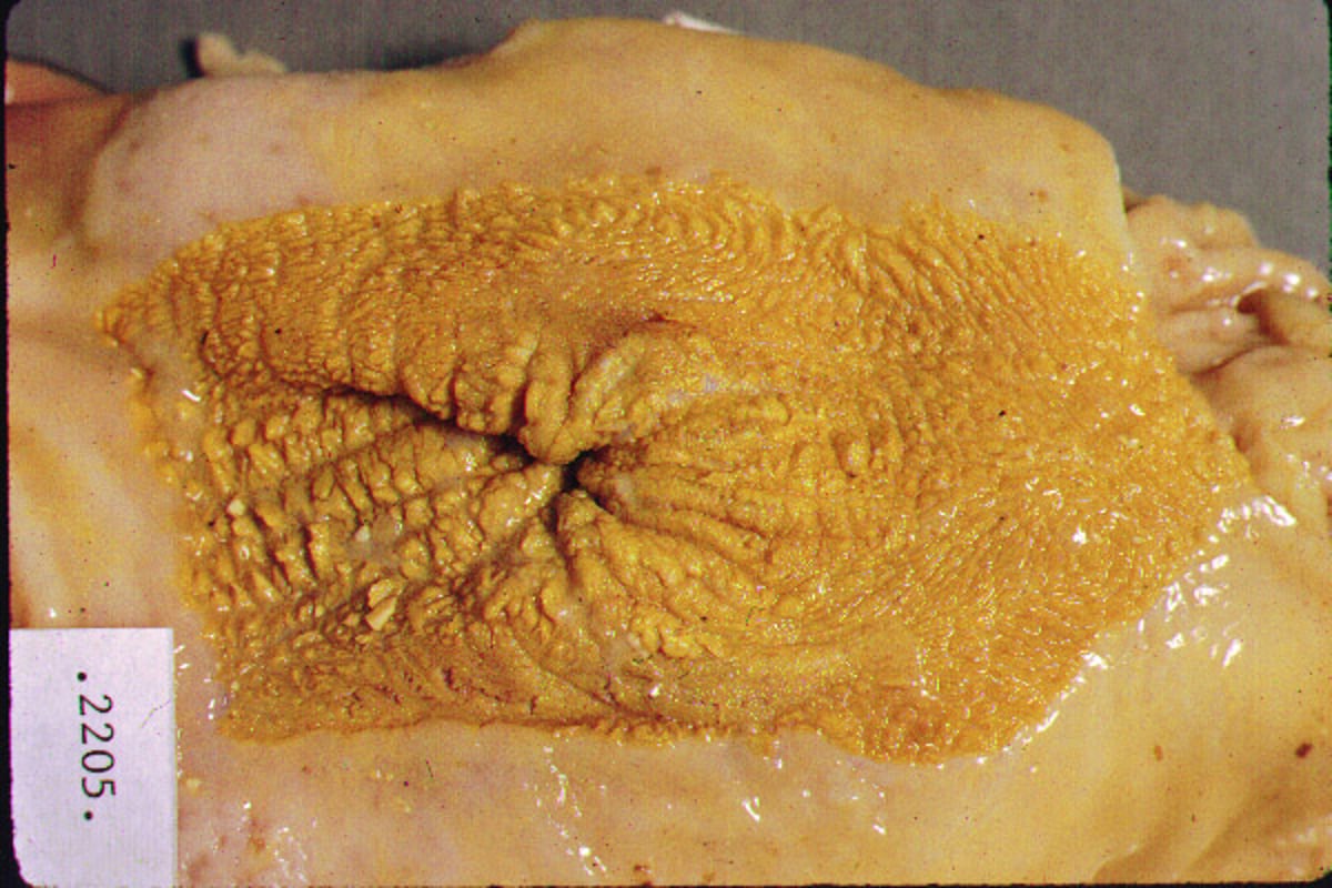
Gastric ulcers in pigs involve the mucosa near the esophageal opening in a rectangular area of white, glistening, nonglandular, squamous epithelium. In a pig that has died suddenly from a gastric ulcer, it is common to find a crater ≥2.5–5 cm in diameter encompassing the distal esophagus. The crater appears as a cream or gray, punched-out area and may contain blood clots or debris. In acute hemorrhage, the stomach and upper small intestine contain dark blood. Earlier lesions are characterized by hyperkeratosis and parakeratosis of the squamous epithelium in the area of the esophageal opening into the stomach. It is assumed that the thickening of the epithelial surface is a defense against insult. Cracks and erosions can occur, particularly near the pars esophageal border. A deep erosion may result in acute and severe blood loss even while most of the surface of the pars esophagea remains intact. The healed ulcer appears as a stellate scar. Erosion and healing may occur over and over. Severe scarring can create stenosis of the distal esophagus.
Diagnosis of Gastric Ulcers in Pigs
Clinical signs including weakness, pallor and sudden death; particularly in the setting of feed disruptions
Erosive lesion(s) located in the pars esophagea and a stomach filled with partially digested blood at necropsy
Appearance in a pen of one or two listless, anorectic pigs that show weight loss, anemia, dark feces, and sometimes dyspnea is suggestive of gastric ulceration, as is the sudden death of an apparently healthy pig. Differential diagnoses include hemorrhagic bowel syndrome, porcine proliferative enteropathy (Lawsonia intracellularis infection), porcine circovirus-associated disease, and swine dysentery. Gross lesions at necropsy are usually sufficient to confirm a diagnosis of gastric ulceration. Abattoir examination has been used to evaluate the prevalence of stomach lesions but must be interpreted with caution. Pigs are generally held without feed before shipping and may be held overnight at the packing plant before slaughter, and during this time ulcers can form or become more severe. On an individual animal basis, possibly for a valuable breeding boar or a pet, an endoscopic examination could readily identify a gastric ulcer.
Treatment and Prevention of Gastric Ulcers in Pigs
Supportive care
Feed changes
No economically feasible treatments are currently available for commercial swine. For pet pigs or valuable breeding animals, proton-pump inhibitors have been shown to reduce gastric acidity and allow mucosal healing. For commercial swine production, weak, pale pigs in poor body condition should be euthanized, and milder cases might respond to palliative care. Placing the affected pig in a hospital pen and initially coaxing it to eat using a highly palatable feed, and possibly administering analgesics, can be attempted. Early marketing of affected pigs should be considered.
Preventing or reducing the incidence of gastric ulcers should focus on maintaining good feed intake, including management steps such as ensuring feeders do not run out of feed, keeping sufficient feeder space available, and minimizing mixing of pigs. During hot weather, it is important to make the pigs as comfortable as possible using cooling methods such as water spraying and plentiful ventilation. Vaccination and other steps to minimize respiratory and systemic diseases are important to prevent gastric ulcer deaths.
Attention to feed milling practices has a major impact on reducing ulcers. Feed produced using a roller mill is regarded to be less ulcerogenic than feed made with a hammer mill. Fine particle size is associated with more likelihood of ulcers but is also associated with improved feed efficiency, so economic considerations must be understood before a drastic change in particle size is recommended. In general, a coarse feed that will protect pigs from stomach lesions may not be an acceptable solution because of the added feed cost, but in an outbreak situation a short-term feed change may be necessary.
Key Points
Gastric ulcers are a common cause of sudden death in grower-finisher pigs and a source of considerable economic loss.
Diagnosis is confirmed at necropsy if the stomach is filled with partially digested blood and the source of bleeding can be identified as deep erosion in the pars esophageal region.
Preventive measures include ensuring the stomach maintains a pH gradient between distal and proximal regions, either by reducing the passage time of feed through the stomach or by encouraging frequent feed consumption.
For More Information
Thomson JR and Friendship RM. Digestive System In: Diseases of Swine, edited by Zimmerman et al. 2019 John Wiley and Sons, Inc. pp 234-263.


