


Infection of the small intestine by type C strains of Clostridium perfringens causes a highly fatal, necrohemorrhagic enteritis. It most commonly affects piglets 1–5 days old; however, in rare cases it occurs in pigs up to 21 days old and other species. Diagnosis is via a combination of lesion assessment and bacterial isolation and typing.
Etiology and Pathogenesis of C perfringens Type C Enteritis in Pigs
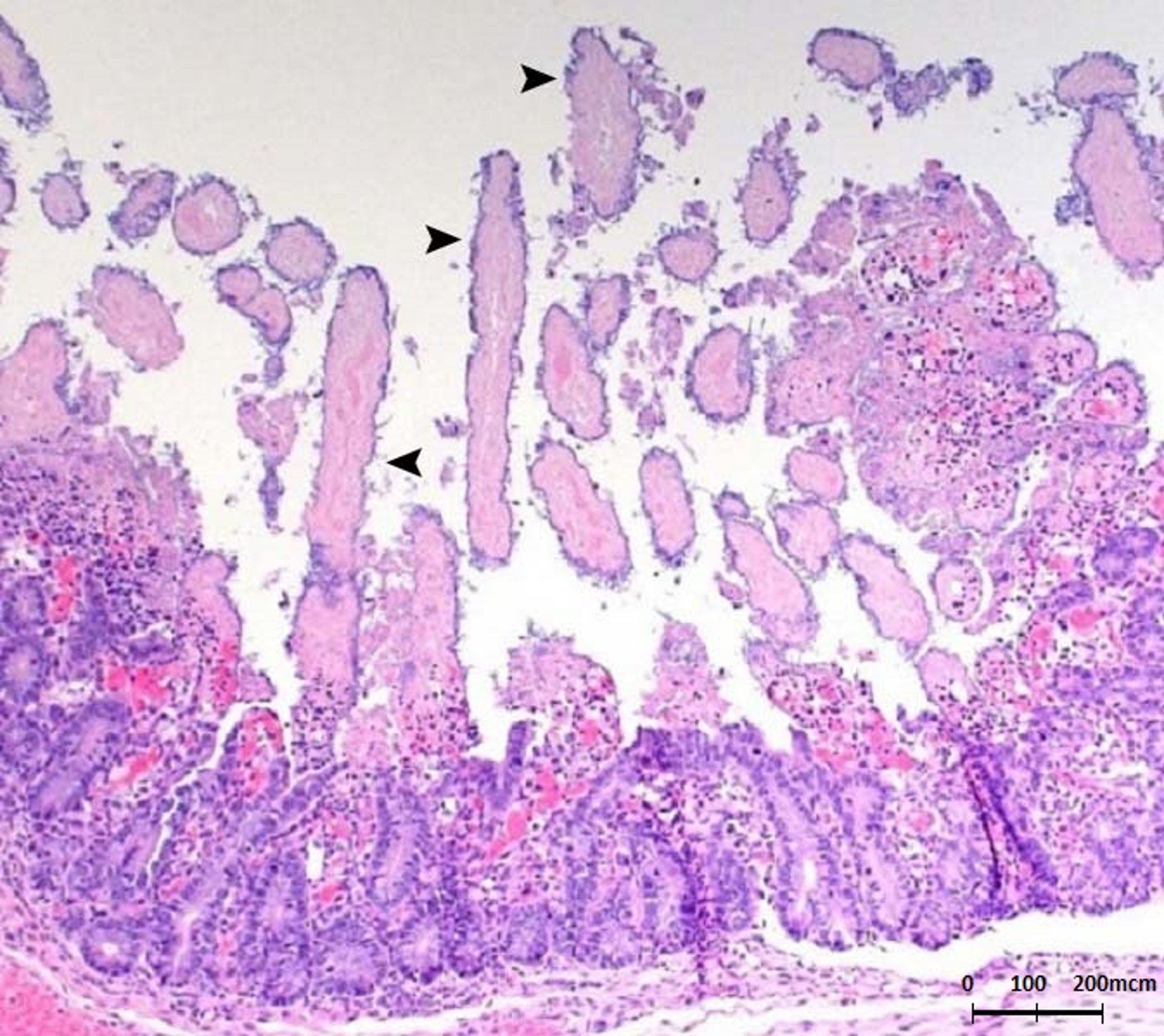
C perfringens proliferates rapidly in the intestinal contents and elaborates beta toxin, a potent, heat-labile, trypsin-sensitive exotoxin that causes necrosis of all structural components of the villi. Necrotizing inflammation usually extends to the mucosal crypts. The infection may continue caudally and involve the ileum; however, it rarely affects the colon. Necrosis of the mucosa is accompanied by blood loss into the intestinal wall and lumen.
Clinical Findings of C perfringens Type C Enteritis in Pigs

Courtesy of Iowa State University Veterinary Diagnostic Laboratory.

Courtesy of Dr. John Prescott.

Courtesy of Dr. Eric R. Burrough.
Sudden onset of hemorrhagic diarrhea followed by collapse and death is characteristic of C perfringens type C enteritis in piglets 1–3 days old. Entire litters are typically affected, and mortality is close to 100%. In less severe cases, brownish liquid feces develop at 3–5 days. Infrequently, pigs develop a persistent, pasty-gray diarrhea and become progressively emaciated.
Lesions
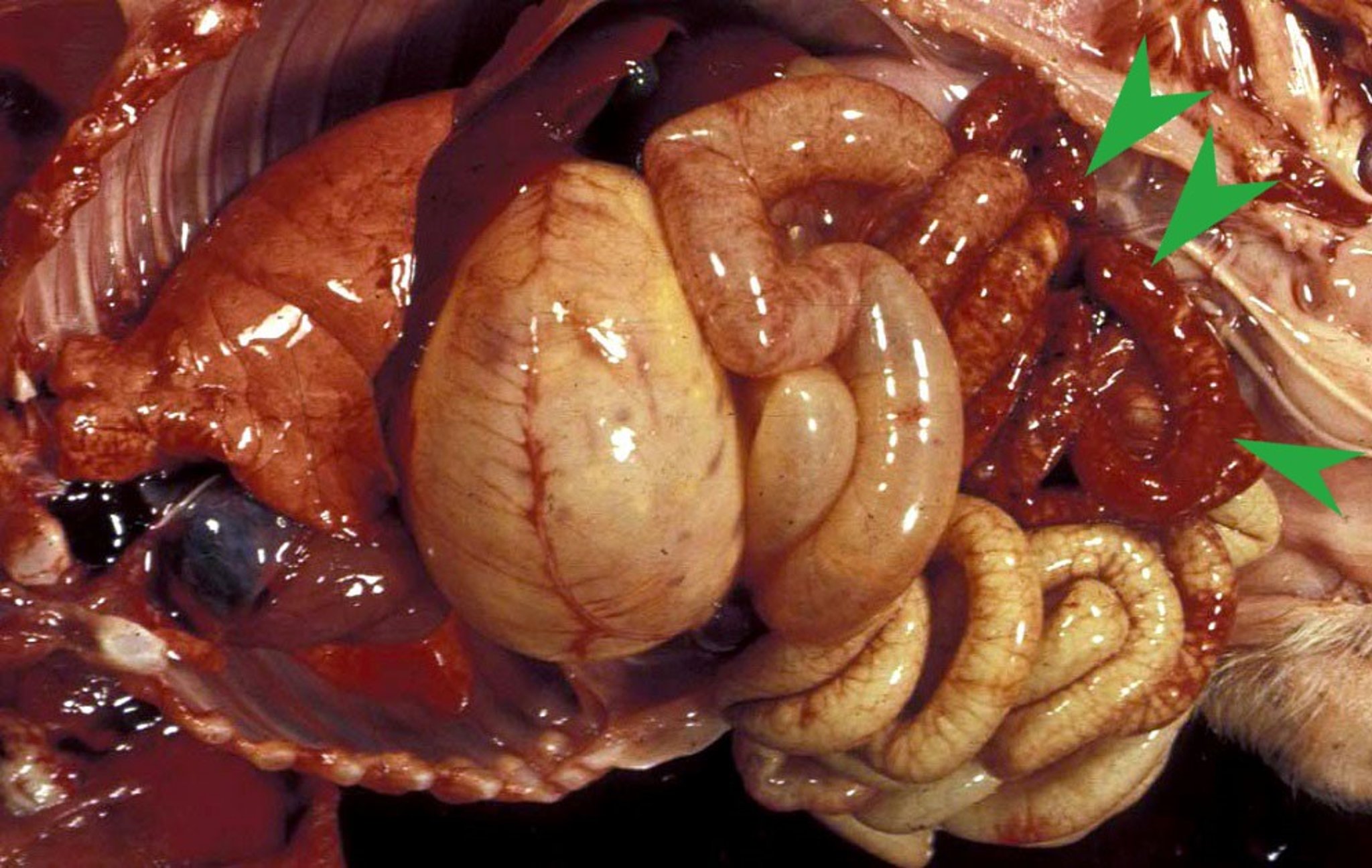
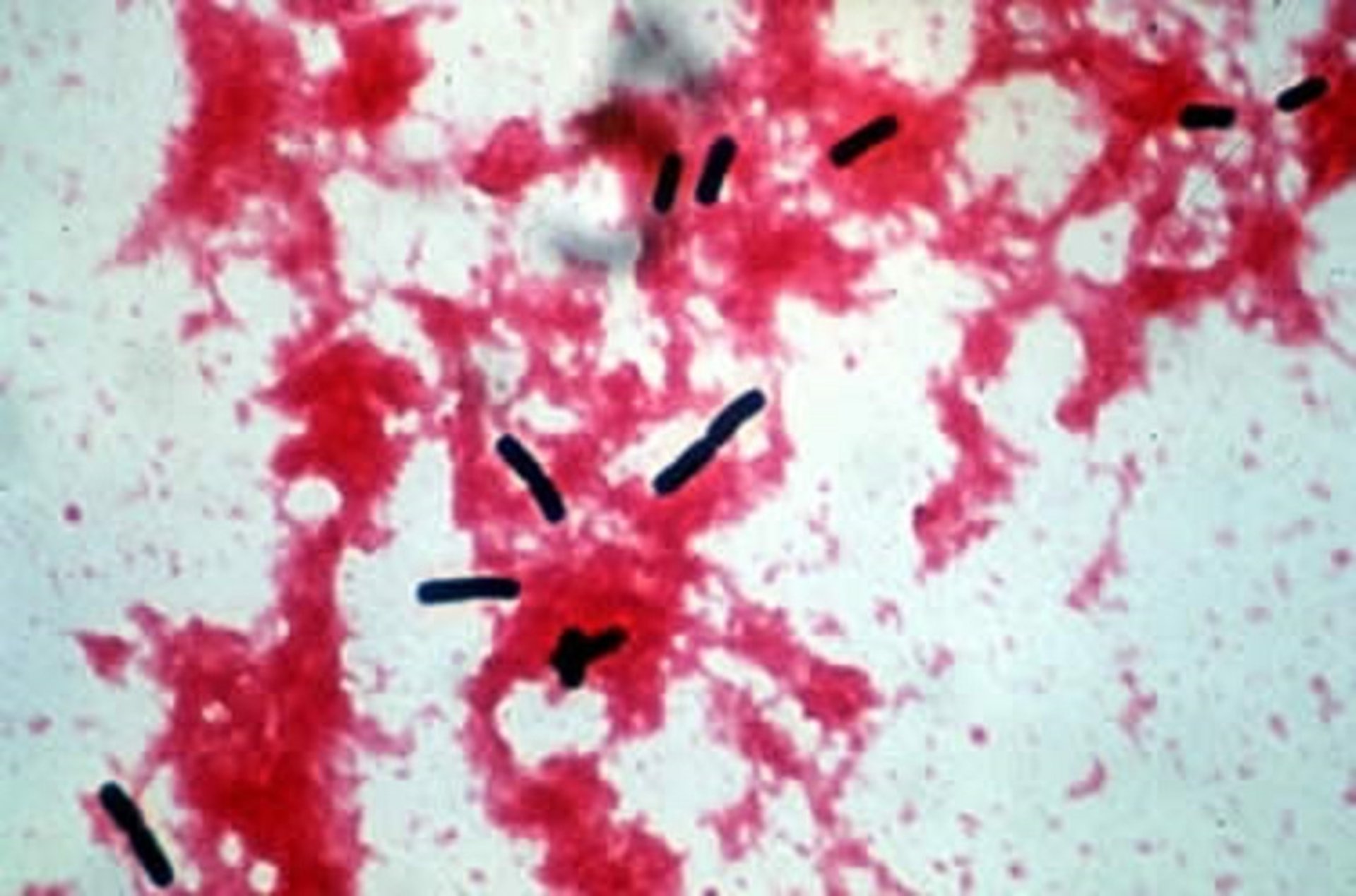
The small intestines of affected piglets are dark red, hemorrhagic, and filled with bloody fluid. At 3–5 days, less severe cases may have emphysema in the wall of the jejunum and necrosis of the mucosa of the jejunum and ileum. More chronic cases have a thickened small intestine lined by a pale yellow or gray necrotic membrane that adheres tightly to the submucosa. Microscopically, there is segmental hemorrhagic necrosis affecting the entire mucosa. Characteristic thick bacilli with square ends are commonly observed along necrotic villi and within the intestinal lumen.
Diagnosis of C perfringens Type C Enteritis in Pigs
Clinical signs and lesions in suckling pigs
Detection of beta toxin in feces, or via culture and genotyping
Postmortem examination is usually sufficient to establish diagnosis of the peracute hemorrhagic form of C perfringens type C enteritis and of the peracute form with jejunal emphysema. A rapid presumptive diagnosis can be made by seeing large, rod-shaped bacteria in Gram-stained mucosal impression smears. Histologic demonstration of villous necrosis with mucosal colonization by numerous large, gram-positive rods is adequate for confirmation. Subacute and chronic forms of the disease in piglets 6–14 days old are easily confused at postmortem examination with Cystoisospora suis enteritis; however, diagnosis is usually possible via histologic examination of the jejunum and ileum or by observation of clostridia in mucosal smears (Gram or Giemsa stain). Isolates of C perfringens may be genotyped for the presence of genes that encode for beta toxin.
Treatment and Control of C perfringens Type C Enteritis in Pigs
Treatment is of little value after clinical signs occur
Increasing lactogenic immunity on sow farms is essential for control
Treatment of pigs with clinical signs of C perfringens type C enteritis is of little benefit because lesions usually are irreversible at the onset of diarrhea. In an acute outbreak, prophylactic administration of type C antitoxin or antimicrobial (or both) parenterally or orally is protective if given to piglets within 2 hours of birth. C perfringens type C enteritis tends to recur on infected premises. Vaccination of gestating sows at 6 and 3 weeks before parturition with type C bacterin-toxoid confers some passive lactogenic immunity if piglets consume colostrum soon after birth. Naive gilts may benefit from three vaccinations before their first farrowing to ensure sufficient colostral antibodies. Once properly immunized, sows should receive one dose ~3 weeks before each subsequent farrowing.
Key Points
C perfringens type C toxin is trypsin-sensitive; thus, disease is common in neonates with low trypsin levels.
Death of entire litters is common.
Epidemics can occur when the organism is introduced into naive herds.
For More Information
Richard OK, et al. Vaccination against Clostridium perfringens type C enteritis in pigs: a field study using an adapted vaccination schedule. Porcine Health Manag. 2019;5:20.
Uzal FA, Songer JG. Clostridial diseases. In Zimmerman JJ, et al, eds. Diseases of Swine, 11th ed. John Wiley and Sons, Inc; 2019:792–806.


