


Enteric colibacillosis is a common disease of nursing and weanling pigs caused by colonization of the small intestine by enterotoxigenic strains of Escherichia coli (ETEC). Colonization of the small intestine by ETEC strains typically results in a severe, watery diarrhea and dehydration. Disease is typically confirmed via a combination of lesion assessment, cultural confirmation, and genotyping of bacterial isolates.
Etiology and Pathogenesis of Enteric Colibacillosis in Pigs

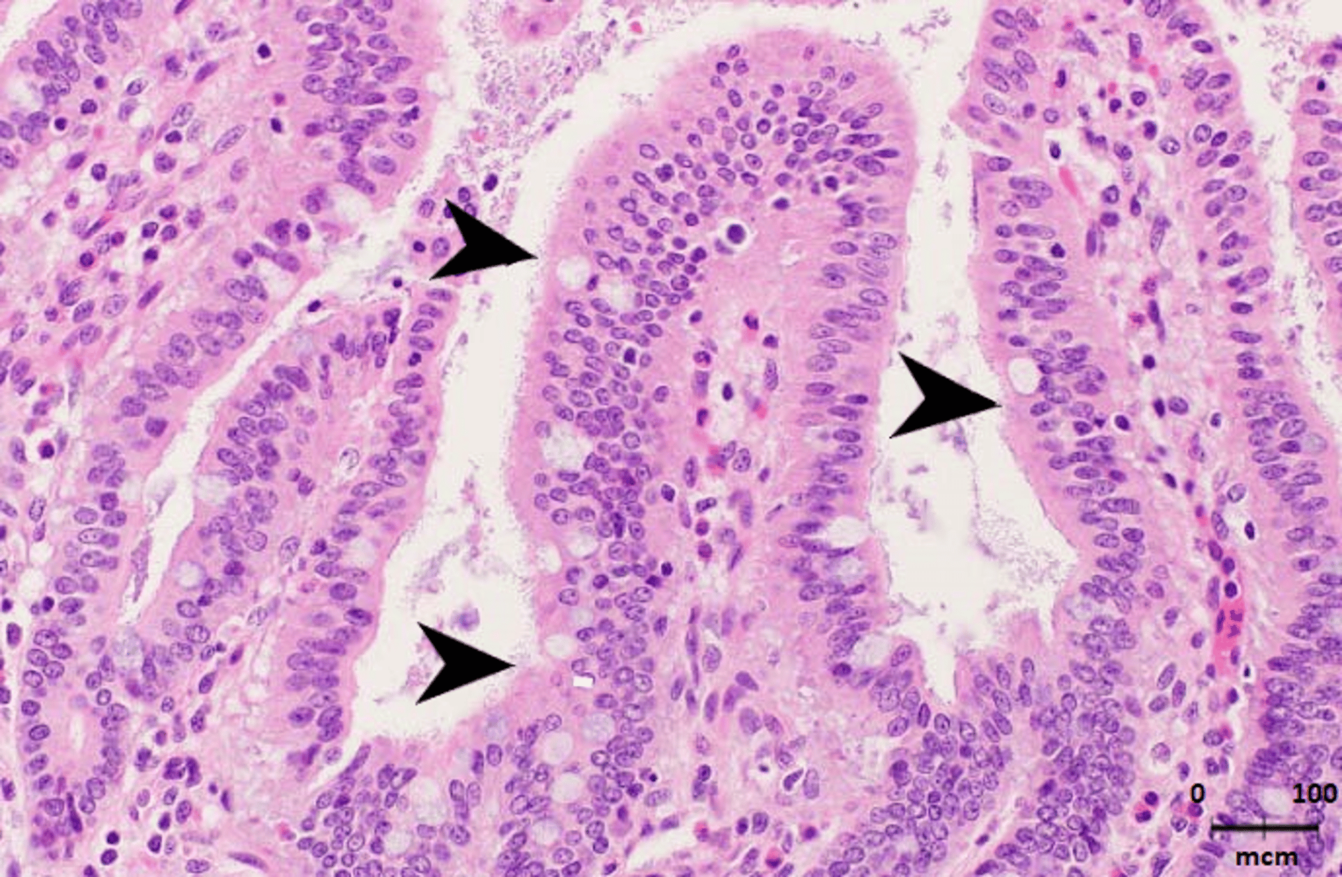
Courtesy of Dr. Eric R. Burrough.
Certain strains of E coli possess fimbriae that allow them to adhere to or colonize the absorptive epithelial cells of the jejunum and ileum. The common antigenic types of fimbriae associated with pathogenicity are F4 (K88), F5 (K99), F6 (987P), F18, and F41. Pathogenic strains produce varying combinations of enterotoxins that act locally to induce fluid and electrolyte secretion into the intestinal lumen, resulting in diarrhea, dehydration, and metabolic acidosis. Neonatal colibacillosis is commonly caused by F4 and, to a lesser extent, F5, F6, and F41 strains. Postweaning colibacillosis is most often associated with the F4 or F18 strain. There is also a genetic susceptibility component, because not all pigs express receptors for all fimbrial adhesins.
Clinical Findings of Enteric Colibacillosis in Pigs
Profuse watery diarrhea with rapid dehydration, acidosis, and death are common clinical signs in pigs with enteric colibacillosis. Rarely, pigs may collapse and die before diarrhea begins.
Lesions
Dehydration and distention of the small intestine, with yellowish, watery to slightly mucoid fluid, is characteristic. The colon often contains similar fluid. The stomach is typically dilated and may be full of feed. Pigs dying suddenly may have patchy cutaneous erythema. Histologically, the villi are usually of normal length and have many small coccobacilli adhered to the absorptive enterocytes.
Diagnosis of Enteric Colibacillosis in Pigs
Confirmation via culture with or without genotyping
Histopathologic evaluation
Confirmation of enteric colibacillosis is based on histologic observation of coliform colonization of small-intestinal brush borders and isolation of the organism from the small intestine. F4 and F18 strains are strongly hemolytic on blood agar; however, not all hemolytic E coli are pathogens. Genotyping PCR assays are available to detect fimbrial type and the presence of enterotoxin genes.
Treatment and Control of Enteric Colibacillosis in Pigs
Antimicrobial treatment based on results of minimum inhibitory concentration testing
Vaccination
Treatment for enteric colibacillosis includes prompt administration of antimicrobials and restoration of fluid and electrolyte balance. Bacterial antimicrobial sensitivity (minimum inhibitory concentration) testing is helpful to identify effective medication, because many strains are resistant to multiple antimicrobials.
Prevention of enteric colibacillosis includes reducing predisposing factors (eg, dampness and cold), improving sanitation, and vaccinating. Gestating sows can be vaccinated with pilus-specific vaccines, and weaned pigs can be treated with live attenuated F4 and F18 strains lacking enterotoxins. Pigs lacking receptors for F4 or F18 E coli are naturally resistant to this disease.
Key Points
Pathogenic E coli have fimbriae for attachment and produce one or more enterotoxins.
Pigs with ETEC develop a secretory diarrhea and dehydrate rapidly.
Because many E coli are commensal organisms, genotyping of isolates is helpful to confirm pathogenic potential.
For More Information
Dubreuil JD, et al. Animal enterotoxigenic Escherichia coli. EcoSal Plus 2016;7(1):10.1128/ecosalplus.ESP-0006-2016.
Fairbrother JM, Nadeau E. Colibacillosis. In: Zimmerman JJ, et al, eds. Diseases of Swine. 11th ed. John Wiley and Sons, Inc; 2019:807–834.
Edema Disease
Edema disease is an acute, highly fatal, neurologic disorder that usually occurs 5 days to 2 weeks after weaning and may be accompanied by diarrhea. See also Edema Disease.


