


Intestinal spirochetosis is a disease limited to the large intestine that commonly manifests as a mucoid diarrhea in grower-finisher pigs. Disease is considerably less severe than what is observed with swine dysentery caused by spirochetes. Intestinal spirochetosis is reported worldwide.
Etiology and Pathogenesis of Intestinal Spirochetosis in Pigs
The etiologic agent of intestinal spirochetosis is Brachyspira pilosicoli. B pilosicoli is emerging as an important pathogen of humans, especially in indigenous populations and immunosuppressed patients. The organism is transmitted orally and survives extremely well in the environment. B pilosicoli has been isolated from a wide variety of animals, including waterbirds, rodents, and dogs. It has been shown to cause diarrheal disease in pigs, chickens, and humans by experimental inoculation and in natural occurrence; however, the pathogenesis is not well studied. As with swine dysentery, B pilosicoli colonization and disease expression can be appreciably influenced by alteration of dietary fiber.
Clinical Findings of Intestinal Spirochetosis in Pigs
Clinical signs of intestinal spirochetosis typically occur in 8- to 12-week-old pigs and often occur 1–2 weeks after movement or commingling. Feces from affected pigs appear similar to wet cement, and this mild diarrhea commonly lasts for 3–6 weeks. Uncomplicated disease is not usually associated with appreciable mortality; however, pigs may have reduced appetite and grow slowly, causing economic losses.
Lesions
The lesions in the large intestine caused by intestinal spirochetosis are milder than those caused by B hyodysenteriae in swine dysentery. The volume of the large intestine may be increased with thickening of the mucosa. In some pigs, a copious catarrhal colitis develops in association with enlarged colonic lymph nodes. Microscopically, spirochetes may be observed attached end-on to the mucosal surface, giving the appearance of a false brush border; however, this lesion is inconsistent and most often occurs early in the course of disease. The mucosal surface may have focal erosions with associated catarrhal exudate. Colonic crypts are often dilated with mucus and contain numerous large spirochetes.
Diagnosis of Intestinal Spirochetosis in Pigs
Selective anaerobic culture for Brachyspira
Histopathologic evaluation

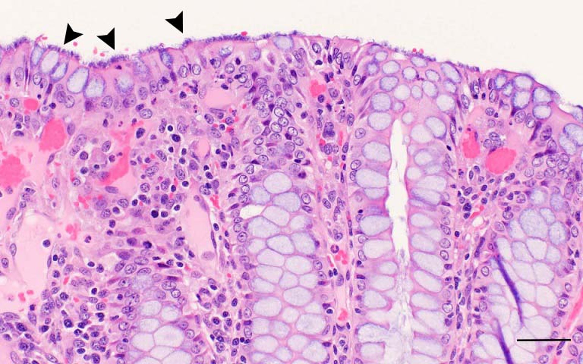
Courtesy of Dr. Eric R. Burrough.
The differential diagnosis of intestinal spirochetosis often includes intestinal salmonellosis, proliferative enteropathy, swine dysentery, and whipworm infection. B pilosicoli and other Brachyspira spp can be isolated on selective agar under anaerobic conditions. Biochemical tests have historically been used for species identification; however, PCR assays, sequencing, and mass spectrometry are increasingly available and more consistent. PCR assays for B pilosicoli that can be applied directly to fecal samples are also available. Histopathologic evaluation with silver stains is useful to identify end-on attached bacteria and to exclude other disease conditions.
Treatment and Control of Intestinal Spirochetosis in Pigs
Antimicrobials
Minimizing exposure to reservoir hosts
Treatment and prevention of intestinal spirochetosis are similar to those of swine dysentery. Administration of drugs such as tiamulin, lincomycin, and carbadox has historically been effective; however, increasing antimicrobial resistance has been reported in several countries, and minimum inhibitory concentration testing is recommended before beginning treatment. It is unknown whether the agent can be eradicated without total depopulation, as for swine dysentery; however, given the number of potential reservoir hosts and environmental survival, it seems unlikely.
Key Points
Disease is limited to the colon and is milder than swine dysentery.
Culture and PCR assays are available for species identification.
Resistance to commonly used antimicrobials is increasing.
For More Information
Hampson DJ. The spirochete Brachyspira pilosicoli, enteric pathogen of animals and humans. Clin Microbiol Rev. 2018;31(1):e00087–17.
Hampson DJ, Burrough ER. Swine dysentery and Brachyspiral colitis. In: Zimmerman JJ, et al,eds. Diseases of Swine, 11th ed. John Wiley and Sons, Inc; 2019:951–970.


