


Determination of the cause of intestinal disease in cattle is based on clinical, epidemiologic, and laboratory findings. Nonspecific therapy includes oral and parenteral fluid therapy to restore the fluid, electrolyte, and acid-base homeostasis. Specific therapy and prevention are detailed under the individual disease headings. Intestinal diseases of neonates are discussed separately, although some of the causes also affect older animals.
Hemorrhagic Bowel Syndrome
Hemorrhagic bowel syndrome (HBS) is a sporadic disease of uncertain etiology that is observed with increasing frequency in cattle. It is manifest by sudden onset of abdominal pain and obstipation progressing to sternal recumbency, shock, and death.
Etiology and Pathogenesis of Hemorrhagic Bowel Syndrome
The etiology of HBS is uncertain but believed to be multifactorial. Clostridium perfringens type A, a normal inhabitant of the bovine digestive tract, has been incriminated as a causative agent, because this organism is isolated from the intestines of naturally occurring cases at a higher frequency and in higher numbers than from cattle with other intestinal diseases.
Another proposed potential causative agent is Aspergillus fumigatus, a common fungus in feed and forages. The primary lesion is similar to that caused by C perfringens in young, rapidly growing animals and consists of an acute, localized, necrotizing, hemorrhagic enteritis of the small intestine that leads to development of an intraluminal blood clot. The clot causes a physical obstruction, with proximal accumulation of intestinal fluid and gas and development of abdominal distension, dehydration, and varying degrees of anemia. The hemorrhagic enteritis is progressive. Ischemia and necrosis extends through the intestinal wall, and, if untreated, there is a fibrinous peritonitis within 24–48 hours, followed by profound toxemia and death.
Epidemiology of Hemorrhagic Bowel Syndrome in Cattle
Hemorrhagic bowel syndrome occurs sporadically, primarily in mature lactating dairy cows in North America and Europe, but it has also been recorded in beef cattle. Most cases occur in mature dairy cattle in the first 3 months of lactation, with highest incidence rates during the cold months of the year. Possible risk factors for disease are those associated with management practices aimed at achieving high milk production, such as high fermentable carbohydrate content of the diet and feeding a total mixed ration. The animal-level disease incidence rate is estimated to be 1%–2%, but outbreaks in a herd can be associated with morbidity rates of ≥10%. Mortality in general is high, with 80%–100% of affected animals dying within 48 hours.
Clinical Findings of Hemorrhagic Bowel Syndrome in Cattle
Cattle affected by hemorrhagic bowel syndrome have a history of:
sudden anorexia and depression
sudden and pronounced drop in milk production
abdominal distention and pain, with kicking at the abdomen
dark, bramble jelly-type feces, scant in amount
dullness and weakness progressing to recumbency
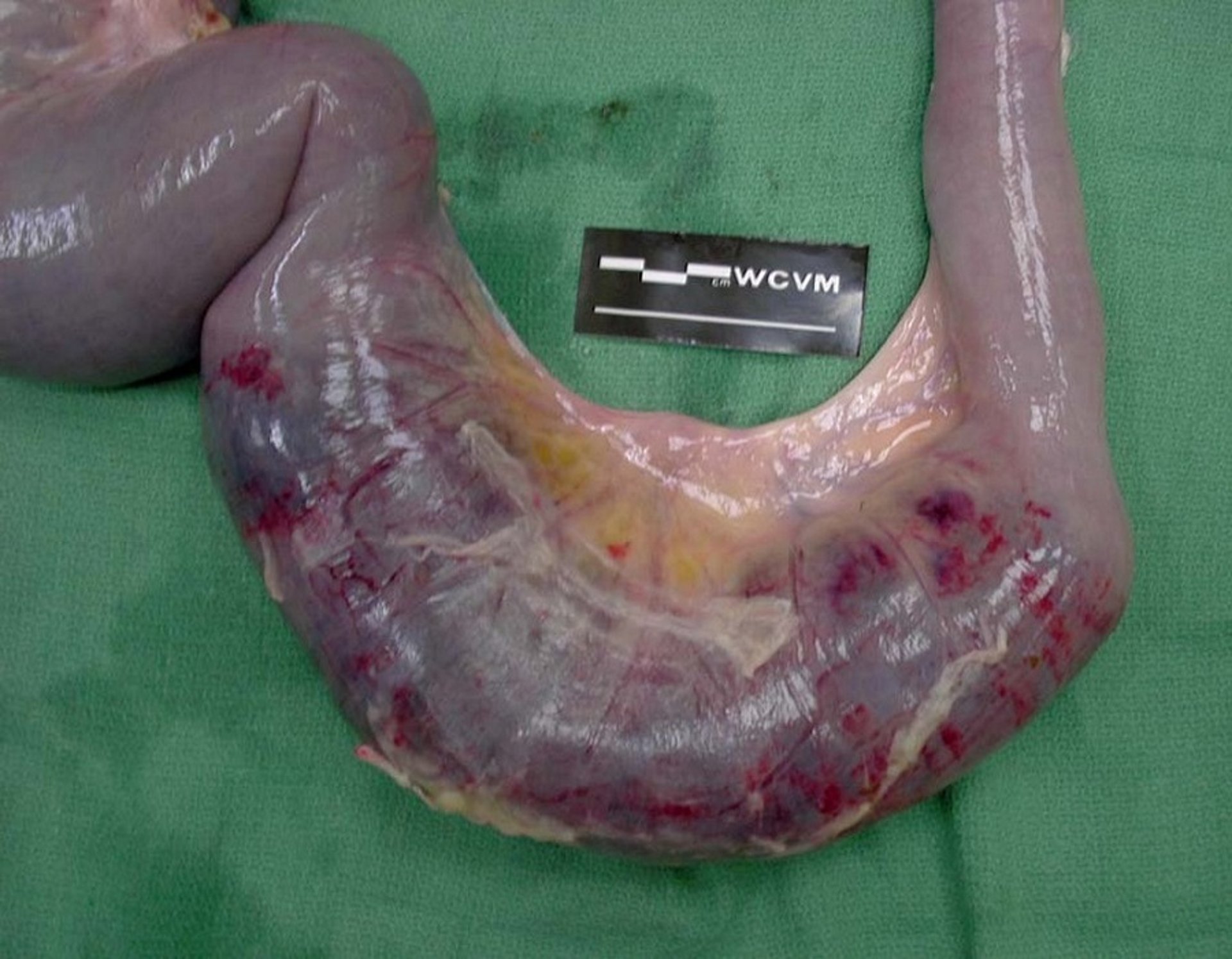
Clinical findings include depression, dehydration, increased heart and respiratory rates, and pale mucous membranes. Abdominal distension of the right flank may be mild but progresses rapidly. Due to the acute onset, there can still be good rumen fill, but the rumen is atonic, and fluid sounds may be elicited by succussion over the right abdomen. Dark red blood clots with a sticky bramble jelly-type consistency are present in the feces. In cases of complete and prolonged intestinal obstruction, the rectum appears dry and sticky, only containing small amounts of dark feces. Distended and firm loops of intestine may be palpable on deep rectal examination. On laparotomy, a segment of the small intestine of variable length is dark red and distended, with a serosal surface covered by tags of fibrin. The small intestine proximal to the affected segment and the abomasum are distended with gas and fluid. Ultrasonography may aid in diagnosis.

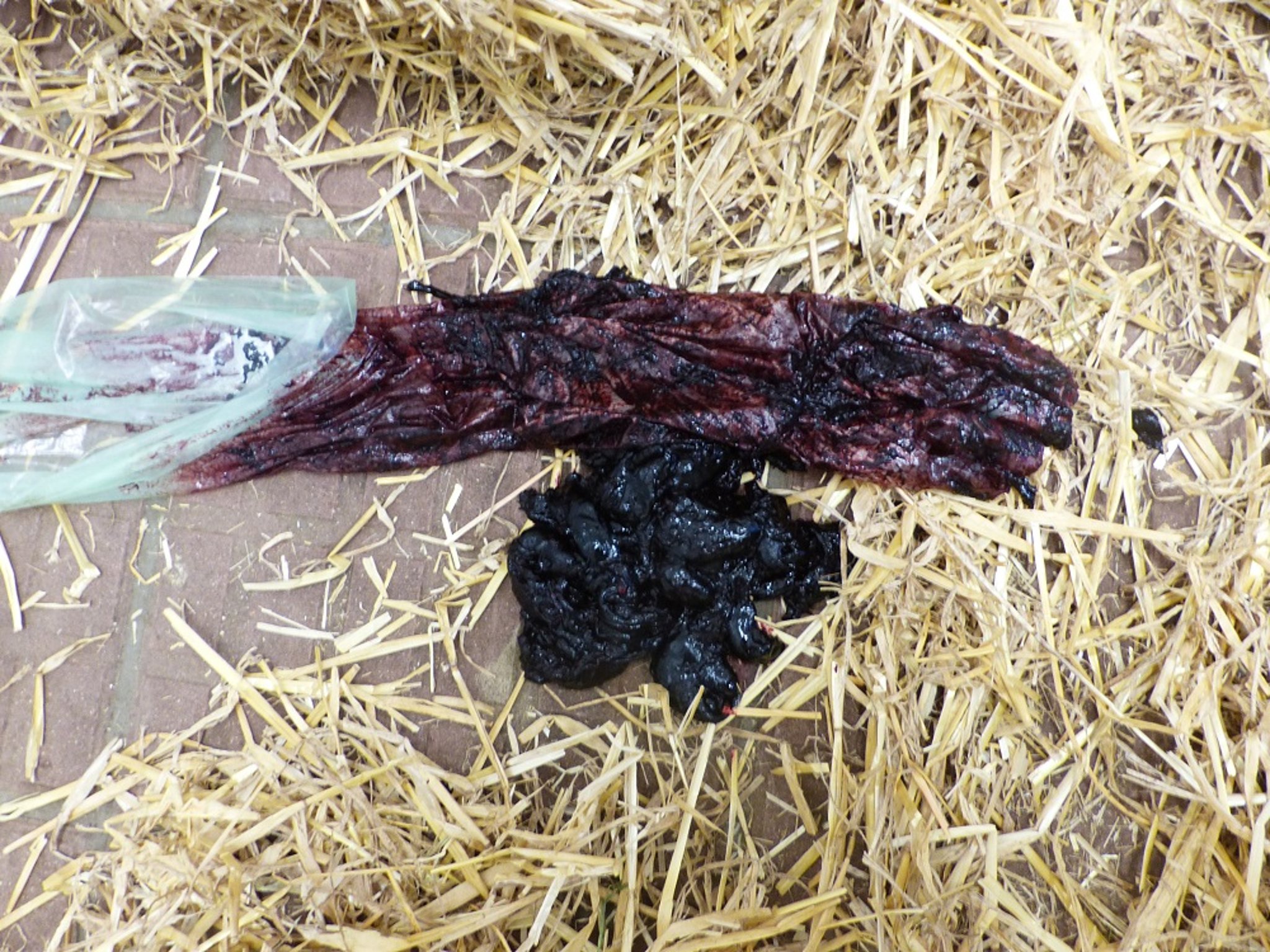
Courtesy of Dr. Walter Grünberg
Most affected cattle die within 2–4 days despite intensive fluid and electrolyte therapy. Sudden death without prior clinical findings may occur. The hemogram is variable; serum biochemistry reflects obstruction of the upper small intestine and sequestration of abomasal secretions with resultant hypokalemia and hypochloremia. Serum L-lactate concentrations are often markedly elevated, with values >5 mmol/L.
Lesions

Courtesy of Dr. Sameeh M. Abutarbush.
Necrohemorrhagic enteritis can occur anywhere in the small intestine and is characterized by severe but localized intraluminal hemorrhage. The affected segment of intestine is dark red and dilated, with tags of fibrin on the serosal surface. The lumen contains a firm blood clot adherent to the mucosa, and at advanced stages the affected segment of intestine becomes necrotic and feels friable on palpation.
Diagnosis of Hemorrhagic Bowel Syndrome in Cattle
Characteristic lesions in the small intestine determined during exploratory laparotomy or at necropsy
A first, tentative diagnosis of hemorrhagic bowel syndrome can be made based on signs of intestinal obstruction in combination with the characteristic appearance of the feces. Diagnosis is confirmed either during an exploratory laparotomy or at necropsy and is based on the presence of a characteristic focal necrohemorrhagic enteritis of the distal small intestine.
Differential diagnoses include:
other causes of physical or functional obstruction of the small intestine (eg, intussusception, cecal dilatation and volvulus)
diffuse peritonitis, from right-sided torsion of the abomasum or torsion at the root of the mesentery
diseases causing melena (eg, abomasal ulcer)
Treatment and Control of Hemorrhagic Bowel Syndrome in Cattle
Surgery and supportive care
Surgery to localize small intestinal loops affected by hemorrhagic bowel syndrome and manually reduce and dislodge blood clots within the intestinal lumen appears to be the most efficient treatment option in the early stages. Fluid and electrolyte therapy is also indicated. In advanced stages, resection of the affected segment of the intestine may be required. Prokinetic substances to enhance intestinal passage administered parenterally combined with single or repeated large, IV doses of heparin to prevent new clot formation in the intestinal lumen may be used postoperatively. The fatality rate is high, and the prognosis is grave. No preventive strategies have been identified. A short-term protective effect of a C perfringens type C and D vaccine against hemorrhagic bowel syndrome in some herds has been reported anecdotally, but there is currently no corroborating scientific evidence.
Winter Dysentery
Winter dysentery is an acute, highly contagious GI disorder that affects housed adult dairy cattle, primarily during winter. Clinical features include profuse diarrhea (sometimes accompanied by dysentery), a profound drop in milk production, variable anorexia and depression, and mild respiratory signs such as coughing. The disease has a high morbidity but low mortality, and spontaneous recovery within a few days is typical.
Etiology of Winter Dysentery in Cattle
Although the precise etiology of winter dysentery has not been conclusively confirmed, an increasing body of evidence implicates a bovine coronavirus (BCoV), closely related to the virus that causes diarrhea in neonatal calves. Evidence for BCoV as the cause of winter dysentery includes the following:
Clinical signs and pathologic findings are consistent with disease induced by BCoV
Seroconversion to BCoV has been demonstrated in affected cattle
The virus is frequently isolated from diarrheic feces of cattle that exhibit clinical signs of winter dysentery
The disease has been reproduced by briefly exposing BCoV-seronegative, lactating cows to a calf experimentally infected with feces from cows with winter dysentery
Notwithstanding, it has not been possible to consistently reproduce winter dysentery through oral inoculation of adult cattle with BCoV. Concurrent risk factors, such as changes in diet, cold temperatures, closed confinement with high animal density, poor ventilation, and presence of other microorganisms, may be required before BCoV causes clinical disease in adult cattle.
Transmission, Epidemiology, and Pathogenesis of Winter Dysentery in Cattle
BCoV is transmitted via the fecal-oral route through ingestion of feed or water contaminated with feces from clinical cases or clinically healthy carrier animals. Viral particles present in respiratory secretions of affected animals may further enhance transmission. Transmission of disease is promoted by close confinement. Winter dysentery is highly contagious and easily introduced to barns by visitors, carrier animals, and fomites. Winter dysentery is common in northern climates where animals are housed indoors for extended periods during the winter months. It is seen frequently in the northern USA, Canada, the UK, Europe, Australia, New Zealand, Israel, and Japan. Coronaviruses survive best at low temperatures and at low ultraviolet light intensities, which can lead to a buildup of virus in the environment during the colder months. Adult lactating cows that have recently calved are most severely affected, but the disease can affect younger or older animals and males.
Mortality rates associated with winter dysentery are generally low (1%–2%), but morbidity in affected herds is high, with 20%–50% of the animals in a herd exhibiting clinical signs within a few days, and close to 100% of animals in the herd exhibiting signs within a week. Some degree of immunity to winter dysentery appears to develop, because recurrences, if seen in the same herd, are noted at 1- to 5-year intervals.
Inflammatory mediators that cause hypersecretion in the small intestine and colon are thought to contribute to the voluminous diarrhea seen in cattle with winter dysentery. In addition, destruction of epithelial cells in the colonic crypts results in transudation of extracellular fluid and blood, explaining the hemorrhagic nature of the diarrhea in some cases.
Clinical Findings of Winter Dysentery in Cattle
Winter dysentery is characterized clinically by an acute onset of fluid diarrhea and a profound decrease in milk production (25%–95% production loss). Feces are liquid and homogenous with little odor, dark green to black, and may contain blood (typically in first-lactation heifers) or mucus. A sweet, musty, unpleasant odor is reported in barns with large numbers of affected cattle. Nasolacrimal discharge or cough may accompany or precede the diarrhea. Other signs include mild colic, dehydration, depression, a brief period of anorexia, and some decrease in body condition. Occasionally, animals exhibit more severe signs such as passage of feces with variable amounts of blood, severe dehydration, and weakness. Fatalities are rare. Diarrhea in individual animals has a short course, and feces return to normal in 2–3 days in most animals. Disease in the herd typically subsides in 1–2 weeks, but milk production may take weeks to months to return to normal.
Lesions
The small intestine may be dilated and flaccid. Lesions are primarily seen in the large intestine and consist of cecal and colonic mucosal hyperemia, linear streaks or pinpoint-sized hemorrhages mostly along the colonic mucosal ridges, and blood in the lumen of the large intestine. Histologic findings may include widespread degeneration and necrosis of colonic glandular epithelium.
Diagnosis of Winter Dysentery in Cattle
PCR and serology
A diagnosis of winter dysentery can be confirmed by demonstrating coronaviral particles in fecal samples via electron microscopy or by confirming the presence of viral antigen or viral DNA via antigen ELISA or reverse transcription/nested PCR, respectively. Seroconversion to coronavirus in acute and convalescent serum samples, taken 8 weeks apart, also helps confirm the diagnosis.
Differential diagnoses for acute diarrhea in adult cattle include bovine viral diarrhea (BVD), enteric salmonellosis, and coccidiosis. These diseases can be excluded by absence of mucosal lesions (BVD), negative fecal cultures (Salmonella spp), and negative fecal flotation (coccidiosis), as well as by the characteristic clinical presentation of winter dysentery (rapid onset of diarrheal disease of short duration in a herd with high morbidity but low mortality).
Treatment and Control of Winter Dysentery in Cattle
Symptomatic and supportive care
Most cattle affected by winter dysentery recover spontaneously. Fresh water, palatable feed, and free-choice salt should be available at all times. The use of astringents, protectants, and adsorbents is controversial. IV fluid therapy or blood transfusions may be required in severely affected cattle.
There is no vaccine for winter dysentery. Isolation of newly introduced cattle for 2 weeks and isolation of any adult cow with diarrhea is advised to decrease the likelihood of disease introduction into a herd. In an outbreak, access to the premises should be restricted, and all persons in contact with affected cattle should ensure that their footwear and clothing are clean before leaving an affected farm.
Other Intestinal Diseases of Cattle
Infection with Salmonella spp can produce diarrhea in animals of all ages, especially those that are stressed, closely stocked, or exposed to a heavily contaminated feed or water supply. In older animals, the disease is manifest by dysentery and toxemia, and mortality can be significant.
Rotavirus and coronavirus occasionally cause outbreaks of diarrhea in suckling calves up to 2–3 months old. The feces are voluminous and may contain mucus. Toxemia is not evident and mortality is negligible, but growth is decreased. (Also see Diarrhea in Neonatal Ruminants.)
Necrotic enteritis of unknown etiology is seen in beef cattle 5–12 weeks old, commonly affecting several calves within the herd. There is sudden onset of fever, depression, and profuse diarrhea. The feces are initially dark green, contain blood, and frequently stain the perineum. Circular erosions may be present in the oral mucosa. A proportion of calves recover after a clinical course of 3–5 days. The clinical course is longer in fatal cases; animals have scant mucohemorrhagic feces that are passed with tenesmus and develop a severe nonregenerative leukopenia. A secondary fibrinous bronchopneumonia may develop. Mortality is high despite intensive antibiotic treatment. At necropsy, there is ulcerative necrosis of the terminal small intestine and the large intestine.
Coccidiosis usually is seen in calves >2 months old but < 1 year old, especially in situations of heavy stocking density and overgrazing. It is characterized by dysentery and tenesmus and may be accompanied by nervous signs. Intestinal helminthiasis, particularly ostertagiosis, is seen in cattle of the same age group. Type I ostertagiosis is seen in cattle on pasture, but Type II ostertagiosis may be seen in housed animals.
Chronic diarrhea and wasting, often in combination with good appetite, occurring as a sporadic disease in adult cattle is typical for paratuberculosis. Chronic diarrhea and wasting also occurring in younger animals may be caused by chronic salmonellosis and chronic BVD infection. Other possible causes of chronic diarrhea include congestive heart failure, uremia, chronic peritonitis, or abdominal mesothelioma. Persistent diarrhea with unthriftiness, and occasionally wasting in yearling and mature cattle, can be associated with a secondary copper deficiency due to excess molybdenum in the pastures. Diarrhea may also accompany selenium-responsive ill-thrift syndromes in growing cattle.
Individual cases or outbreaks of diarrhea may be associated with dietary indiscretions. Diarrhea may follow cases of simple indigestion and is common in grain overload. It also follows ingestion of toxic amounts of chemicals (eg, arsenic, copper, zinc, and molybdenum) or certain poisonous plants and mycotoxicoses; dipyridyl and organophosphate poisoning can also cause diarrhea.
Cattle may also harbor organisms such as Escherichia coli O157:H7, Yersinia enterocolitica, and Campylobacter jejuni in the intestine. Although these are rarely associated with clinical disease in cattle, fecal contamination of milk may lead to outbreaks of gastroenteritis in people who consume unpasteurized milk or cheese products. Retail meat products can also be infected if there has been fecal contamination of the carcass at slaughter.
Intestinal adenocarcinoma, commonly seen in association with bovine enzootic hematuria, is believed to result from the interaction of a carcinogen (ptaquiloside) in bracken fern (Pteridium spp) and papillomavirus.
Intestinal obstructions are seen sporadically. Cecal dilatation and volvulus are seen predominantly in adult cattle in the postparturient period. Intussusception occurring at the distal jejunum or proximal ileum is the most common cause of complete obstruction in both adult cattle and calves. Ileocecocolic, cecocolic, and colonic intussusceptions are seen less frequently in calves and not at all in adult cattle because of the greater strength of the ileocecal ligament and the presence of mesenteric fat, which stabilize this region of the bowel in older cattle. Intestinal volvulus and volvulus around the mesenteric root are seen sporadically at all ages. Rarely, intestinal obstruction is caused by incarceration and entrapment of the small intestine by persistent urachal or umbilical remnants, by obstruction of the small intestine or descending colon by phytobezoars and enteroliths, or by compression from fat necrosis or lipoma.
Intestinal obstruction can also be caused by congenital disease, most commonly by atresia coli (which is seen both sporadically and in clusters on a farm and may be caused by rectal palpation of the amniotic vesicle at 35 and 41 days of pregnancy) but also by atresia ani (which may be accompanied by urogenital defects and defects of the tail).
A congenital disease affecting endogenous cholesterol transport has recently been described in Holstein Friesian cattle and is recognized as cholesterol deficiency haplotype. The disease is characterized by progressive emaciation and incurable diarrhea over the first months of life, while affected calves maintain a vigorous appetite. Blood cholesterol levels are persistently at or below the detection limit. Homozygous carriers of the gene defect typically die of emaciation in the first year of life. Heterozygous carriers only show moderately decreased blood cholesterol concentrations but no obvious clinical signs.


