


Cushing disease is hyperadrenocorticism caused by an ACTH-secreting tumor of the pituitary gland. Clinical signs include polyuria, polydipsia, alopecia, and muscle weakness. A low-dose dexamethasone suppression test is the preferred diagnostic test. Treatment options include medical therapy, radiation, and surgery.
Cushing disease, or pituitary-dependent hyperadrenocorticism, arises from adenomatous enlargement of the pituitary gland, resulting in excessive ACTH production. A related term, Cushing syndrome, refers to elevated adrenocortical hormones, regardless of cause. The latter term includes pituitary-dependent hyperadrenocorticism as well as adrenal-dependent disease, which is associated with functional adenomas or adenocarcinomas of the adrenal gland. Ectopic ACTH secretion has not been reported in dogs; however, in people, ectopic ACTH secretion is associated with certain lung tumors. Iatrogenic hyperadrenocorticism results from chronic excessive exogenous steroid administration.
Clinical Findings of Cushing Disease in Animals
Cushing disease is seen in middle-aged to older dogs (7–12 yr old); ~85% have pituitary-dependent hyperadrenocorticism (PDH), and ~15% have adrenal tumors.
Breeds in which Cushing disease is commonly seen include:
Poodles, especially miniature poodles
Dachshunds
Boxers
Boston terriers
Yorkshire terriers
Staffordshire terriers
Large-breed dogs often have adrenal tumors, and there is a distinct predilection in females (3:1). In cats, hyperadrenocorticism is found in middle-aged to older cats, with a slight predilection in females (60%).

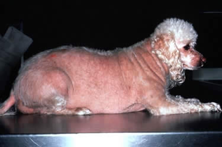
Courtesy of Dr. Stephen White.
Common clinical signs of Cushing disease in dogs and cats are:
polydipsia
polyuria
polyphagia
heat intolerance
lethargy
abdominal enlargement ("pot-belly")
panting (dogs only)
muscle weakness
recurrent urinary tract infections
alopecia
thin, fragile skin (especially cats)
phlebectasias
comedones
bruising
cutaneous hyperpigmentation
calcinosis cutis
pyoderma
dermal atrophy (especially around scars)
secondary demodicosis
seborrhea
In cats, the most striking dermatologic sign is increased skin fragility; many cats present with self-inflicted cutaneous wounds. Secondary infections (especially respiratory) are also common in cats.
Uncommon clinical manifestations of Cushing disease include:
hypertension
pulmonary thromboembolism
bronchial calcification
congestive heart failure
polyneuropathies
polymyopathies
behavioral changes
blindness (central)
pseudomyotonia
nonhealing corneal ulceration
cranial cruciate ligament rupture
perianal adenoma in a female or castrated male dog
clitoral hypertrophy
testicular atrophy in intact males
penile barbs in castrated male cats
prostatomegaly in castrated male dogs
In dogs, serum chemistry abnormalities associated with hypercortisolemia include:
increased serum alkaline phosphatase (SAP)
increased ALT
hypercholesterolemia
hyperglycemia
decreased BUN
Hypercholesterolemia is due to steroid stimulation of lipolysis. SAP is increased primarily from induction of a specific hepatic isoenzyme; some also comes from hepatic glycogen deposition and vacuolization impinging on the biliary system. SAP is seldom increased in cats (< 20%), because they lack the specific hepatic isoenzyme for it. Increased serum ALT and AST are caused by hepatocellular necrosis, glycogen accumulation, and swollen hepatocytes. Decreased serum phosphorus may be a result of increased urinary excretion due to polyuria. Abnormalities noted on the biochemical profile may also include hyperglycemia due to increased gluconeogenesis and decreased peripheral tissue utilization through insulin antagonists. Approximately 10% of Cushingoid dogs are diabetic; however, in cats with hyperadrenocorticism, almost 80% present with overt diabetes mellitus and insulin resistance.
The hemogram is characterized by evidence of regeneration (erythrocytosis, nucleated RBCs) and a classic stress leukogram (eosinopenia, lymphopenia, and mature leukocytosis). Basophilia is occasionally seen. Many dogs with hyperadrenocorticism show evidence of urinary tract infection without pyuria (positive culture), bacteriuria, and proteinuria resulting from glomerulosclerosis. In cats, polydipsia and polyuria are a result of concurrent diabetes mellitus, and urine specific gravity is usually high. In dogs, cortisol-induced interference with ADH binding results in hyposthenuria, and central diabetes insipidus may occur as a result of pituitary tumor enlargement.
Diagnosis of Cushing Disease in Animals
Low-dose dexamethasone suppression test
There is no single test or combination of tests that is 100% accurate to diagnose Cushing disease. The sensitivity and specificity of individual tests or combinations of tests are increased when they are applied to a patient population likely to have hyperadrenocorticism. The diagnosis should be based on appropriate clinical signs followed by supporting minimum database abnormalities (eg, high cholesterol, SAP), and confirmed via an appropriate screening test for hyperadrenocorticism. If screening test results are inconclusive, or if laboratory abnormalities associated with hyperadrenocorticism (eg, increased SAP) are noted in a dog without clinical signs, the dog should be retested 3–6 mo later rather than treated without a definitive diagnosis. In particular, the diagnosis of sex steroid–induced Cushing disease may be especially difficult.
The urine cortisol to creatinine ratio (UCCR) is a highly sensitive test to differentiate healthy dogs from those with hyperadrenocorticism, but it is not highly specific because dogs with moderate to severe nonadrenal illness also exhibit increased ratios. UCCR should be determined based on free-catch urine collected at home by the owner. The stress of transporting a dog or cat to the veterinary hospital, the stress of cystocentesis, or both, can be enough to cause a falsely increased UCCR. An increased UCCR should be confirmed with an ACTH stimulation test, an IV low-dose dexamethasone suppression (LDDS) test, or an oral LDDS test.
The LDDS test is the screening test of choice for canine hyperadrenocorticism when properly used. Only 5%–8% of dogs with PDH exhibit suppressed cortisol concentrations at 8 hours (ie, are false-negatives). In addition, 30% of dogs with PDH exhibit suppression at 3 or 4 hours followed by “escape” of suppression at 8 hours; this pattern is diagnostic for PDH, making further testing unnecessary. The major disadvantage of the LDDS test is the lack of specificity in dogs with nonadrenal illness: >50% of dogs with nonadrenal illness have a positive LDDS test. In such cases, the dog should be allowed to recover from the nonadrenal illness before testing for hyperadrenocorticism with an LDDS test.
Another option, particularly in cats, is to perform an oral LDDS test using the UCCR as the discriminator. In this test, morning urine is collected for a baseline on days 1 and 2. On the second day after urine collection, three doses of dexamethasone at 0.1 mg/kg (cats) or 0.01 mg/kg (dogs) is administered every 6 hours, and a urine sample is collected for UCCR measurement the next morning. After analysis of the first two UCCRs have shown elevation in urine cortisol to confirm the diagnosis of hyperadrenocorticism, the two samples can be combined into a single "pre" or baseline sample. If the UCCR decreases by 50% after oral dexamethasone, the diagnosis of pituitary-dependent hyperadrenocorticism can be confirmed However, a diagnosis of adrenal-dependent hyperadrenocorticism cannot be assumed by the lack of suppression; therefore, further testing using endogenous ACTH or imaging via ultrasound may be needed.
The ACTH stimulation test is used to diagnose various adrenopathic disorders, including endogenous or iatrogenic hyperadrenocorticism and spontaneous hyperadrenocorticism. As a screening test for the diagnosis of naturally occurring hyperadrenocorticism, it has a diagnostic sensitivity of ~80%–85% and a higher specificity than the LDDS test. In one study, only 15% of dogs with nonadrenal disease had an exaggerated response to ACTH stimulation. Adrenal tumors may be particularly difficult to diagnose using an ACTH stimulation test; however, an ACTH stimulation test is the test of choice for iatrogenic hyperadrenocorticism.
Dogs with adrenal sex steroid excess may have negative ACTH stimulation and LDDS tests, because serum cortisol concentrations are normal. This may be due to excess cortisol precursors. Increases in progesterone, 17-OH-progesterone, androstenedione, testosterone, and estrogens may require dynamic adrenal testing using the ACTH stimulation test and measurement of sex steroids in addition to cortisol.
After the diagnosis of hyperadrenocorticism has been confirmed, differentiation of pituitary- versus adrenal-dependent disease may be necessary. Although most dogs with hyperadrenocorticism have PDH, in atypical cases (eg, anorectic dogs with hyperadrenocorticism), a differentiation test is appropriate. In particular, differentiation of PDH (often macroadenomas) from adrenal tumors is often necessary in large breeds.
The high-dose dexamethasone suppression (HDDS) test works on the principle that autonomous ACTH hypersecretion by the pituitary can be suppressed by supraphysiologic concentrations of steroid. Dogs with autonomous cortisol-producing adrenal tumors have maximally suppressed ACTH production via the normal feedback mechanism; therefore, administration of dexamethasone, no matter how high the dose, cannot suppress serum cortisol concentrations. In dogs with PDH, however, the high dose of dexamethasone is able to suppress ACTH and, hence, cortisol secretion. One important caveat is that dogs with pituitary macroadenomas (15%–50% of dogs with PDH) do not suppress ACTH on the HDDS test.
Measurement of endogenous plasma ACTH concentrations is the most reliable way to discriminate between PDH and adrenal tumors. Dogs with adrenal tumors have low to undetectable ACTH concentrations; in contrast, dogs with PDH have normal to increased ACTH concentrations. Recently, researchers have found that the addition of the protease inhibitor, aprotinin, to whole blood in EDTA tubes inhibits the degradation of ACTH. Samples may be collected, spun in a nonrefrigerated centrifuge, and kept for as long as 4 days at < 4ºC.
Diagnostic imaging of the pituitary and the adrenal glands can be accomplished via abdominal radiography, ultrasonography, CT, or MRI. Abdominal radiographs should be obtained for all dogs that do not suppress on an HDDS; ~30%–50% of dogs with adrenal tumors have a mineralized mass in the area of the adrenal glands. Abdominal ultrasonography is a more sensitive way to identify adrenal tumors. In addition, liver metastasis or invasion into the vena cava may be demonstrated in dogs with adrenal carcinomas. CT or MRI of the brain or abdominal cavity in dogs that do not suppress on the HDDS may demonstrate unilateral adrenal enlargement (50%), pituitary macroadenoma (25%), or pituitary microadenoma (25%).
Treatment and Prognosis for Cushing Disease in Animals
Medical, surgical, and radiation therapy
Three treatment options are available for Cushing disease in dogs. Medical, surgical, and radiation therapy have all been used with varying degrees of success.
Dogs with PDH may be treated using the adrenolytic agent mitotane (o,p′-DDD), beginning with an induction dosage of 25–50 mg/kg/day for 7–10 days. Dogs should be monitored for signs of hypoadrenocorticism, such as anorexia, vomiting, and diarrhea; if such signs occur, mitotane therapy should be discontinued and glucocorticoids administered. Water consumption or appetite may be measured to provide an endpoint for therapy; water consumption should decrease to < 60 mL/kg/day (dogs). Appetite is a more precise way to monitor mitotane therapy in many cases. To monitor therapy, the dog is fed 75-80% of its normal ration, and when the dog doesn't finish a meal the owner should bring the dog in for ACTH response testing. After 7–10 days of therapy with mitotane or a reduction in water or food consumption, an ACTH response test should be performed to determine whether cortisol suppression is adequate. Cortisol levels measured both before and after the ACTH response test should be in the normal range. To maintain suppression of cortisol secretion, mitotane is administered at a dosage of 50 mg/kg per week. Dogs on longterm treatment with mitotane should have an examination and ACTH response test every 3–4 months. Gradually increasing dosages of the drug are often required to maintain adequate clinical remission.
Adverse effects of mitotane at the recommended dosage include GI irritation (vomiting and anorexia), CNS disturbances (ataxia, weakness, seizures), mild hypoglycemia, and a moderate increase in SAP. Signs such as depression or ataxia can be alleviated by dividing the daily dose into two equal parts administered at 8- to 12-hour intervals. Persistence of CNS signs after mitotane is discontinued suggests an expanding pituitary macroadenoma.
Reports have demonstrated the efficacy of the adrenal enzyme inhibitor trilostane in the treatment of PDH in dogs. Studies in dogs with hyperadrenocorticism have shown that trilostane is an effective steroid inhibitor with minimal adverse effects. Trilostane should be administered every 12 hours at a starting dose of 1–3 mg/kg, PO (given with food), to achieve a decrease in glucocorticoid secretion from the adrenal glands. Mineralocorticoid insufficiency, which is reversible, can also be seen in animals receiving trilostane; a few cases of adrenal necrosis with permanent adrenal insufficiency have been seen after trilostane administration. Trilostane may prove to be a reasonable alternative to mitotane therapy for PDH in dogs. Monitoring of trilostane treatment can be tricky; however, most people use the ACTH stimulation test to maintain post ACTH cortisol levels in the normal range. Once-daily therapy with trilostane can be associated with transient hypoadrenocorticism, which may be difficult to identify with routine testing. Another option for monitoring is to determine pre-trilostane or 3-hour post trilostane cortisol concentrations. Pre-trilostane and 3-hour post trilostane cortisol concentrations ≤ 138 nmol/L or 62 nmol/L, respectively, were associated with excellent control based on clinical signs observed by the owner.
Radiation therapy of pituitary tumors is associated with a high rate of response; however, most dogs and cats require ancillary trilostane or mitotane therapy for several months after radiation treatment because of residual ACTH secretion. In dogs with PDH undergoing hypophysectomy, 80% achieved remission, with an 11% recurrence; thyroid and glucocorticoid support may be needed after surgery, and animals may lose the ability to secrete vasopressin, leading to diabetes insipidus as well. Selegiline is an irreversible monoamine oxidase (type B) inhibitor that increases dopamine levels. Dopamine inhibits ACTH release from the pituitary gland. However, only ~20% of cases with PDH can be expected to respond; no significant changes in serum cortisol or creatinine, ACTH stimulation, or LDDS have been noted with selegiline therapy.
Treatment of iatrogenic hyperadrenocorticism should include a change to an oral, short-acting steroid such as prednisone or prednisolone. Gradually, the steroid dose is decreased from ~1 mg/kg to 0.5 mg/kg throughout several weeks and then tapered to an alternate-day schedule until the adrenal glands can respond to ACTH stimulation. Monthly ACTH stimulation tests may be performed to determine when steroid treatment can be discontinued.
Surgical removal of unilateral adrenal adenomas or adenocarcinomas may be indicated in some cases; however, surgical and anesthetic complications (eg, hypotension) may develop secondary to hypoadrenocorticism, which occurs immediately after surgical removal of the tumor. Median survival for dogs with carcinomas treated with surgical excision was 778 days. The metastatic rate was 5% at time of surgery and 14% longterm. With unilateral adrenalectomies, mortality within 1 month after surgery was 14%–60%; overall rate for cure for adrenal tumors was ~50%. Medical treatment of adrenal tumors is difficult because they tend to be resistant to the effects of mitotane. Adrenal tumors are relatively resistant to mitotane; dogs require as much as four times the dose of mitotane to respond, and clinical response tends to be less favorable.
Finally, if the dog is showing neurologic signs (eg, anorexia, stupor, or seizures) and a large pituitary tumor (macroadenoma) is identified, radiation therapy of the pituitary gland is indicated. Newer types of radiation therapy (cyberknife, gamma knife) may prove to be superior to previously available modalities and can treat pituitary tumors in < 3 days with minimal adverse effects. Results of radiation therapy in dogs show that this is an effective method of treatment with low morbidity; however, it may take several months for the signs of PDH to subside. These dogs do well longterm, however, because the primary disease process (pituitary tumor) has been addressed. Hypophysectomy is available on a limited basis and has many advantages over conventional therapies, such as rapid reversal of clinical signs and excellent prognosis. Side effects of hypophysectomy can include transient diabetes insipidus, infection, and complications arising from rapid reversal of the hyperadrenocorticism.
Prognosis of dogs with PDH has been estimated to be ~2 years with or without medical therapy. Radiation treatment of pituitary tumors causing PDH or hypophysectomies is associated with a relatively good longterm prognosis (2–5 years). Prognosis of dogs with unilateral adrenalectomy was 18 months.
Key Points
Cushing disease, or pituitary hyperadrenocorticism, is a common endocrine disorder of dogs and a rare disorder of cats.
Clinical signs of Cushing disease, such as polydipsia or polyuria, are similar between cats and dogs, with the exception that cats are more likely to have concurrent diabetes mellitus and skin fragility.
Treatment of Cushing disease in dogs include medical therapy (mitotane, trilostane), radiation therapy, and surgery (hypophysectomy)
For More Information
Also see Pet Health content regarding Cushing disease in dogs and Cushing disease in cats.


