


True acanthosis nigricans is a genetic dermatosis found primarily in Dachshunds. Postinflammatory hyperpigmentation may appear clinically similar, but is a secondary reaction following an underlying disorder. Diagnosis is made based on the clinical presentation. Treatment includes topical anti-inflammatory drugs or antimicrobials if secondary bacterial or yeast infection is present.
Acanthosis nigricans has two presentations. The most common type is a clinical reaction due to inflammation in dogs characterized by axillary and/or inguinal hyperpigmentation, lichenification, and alopecia. The second is a genodermatosis primarily of Dachshunds that occurs early in life and is noninflammatory. Currently, dermatologists do not use the term acanthosis nigricans to describe the inflammatory reaction pattern. The challenge is identification of the underlying skin disease that is triggering the reaction pattern.
Etiology and Clinical Findings of Acanthosis Nigricans
Acanthosis nigricans is a disorder of hyperpigmentation. There is no sex predilection. Primary acanthosis nigricans is a genodermatosis that can occur in many breeds, but particularly Dachshunds. Clinical signs are usually present by 1 year of age in this breed.
Secondary acanthosis nigricans is really postinflammatory hyperpigmentation and should be referred to as such, ie, the term "acanthosis nigricans" is not appropriate. This inflammatory reaction pattern can occur in any breed of dog and at any age; it is most common in breeds predisposed to conditions that result in inflammation of the axillary or inguinal region due to:
conformational abnormalities
obesity
endocrinopathies (eg, hypothyroidism, hyperadrenocorticism, sex hormone abnormalities)
axillary and inguinal pruritus associated with atopic dermatitis
food allergy
contact dermatitis
primary disorders of keratinization
skin infections (eg, staphylococcal pyoderma, Malassezia dermatitis)
Clinical signs start with increased pigmentation in the axillary and/or inguinal region. In primary acanthosis nigricans, the hyperpigmentation is initially diffuse and noninflammatory. It tends to develop uniformly in the affected areas. In primary acanthosis nigricans, secondary inflammatory lesions (ie, lichenification) most commonly develop as a result of conformational friction.
In postinflammatory hyperpigmentation, the distribution is patchy and often starts with a lacy appearance. It may not develop in all areas at the same time. Inflammation is mild but becomes more severe with time. Lesions of postsecondary inflammation are not necessarily present in both the axillary and inguinal region, nor are they necessarily symmetric.

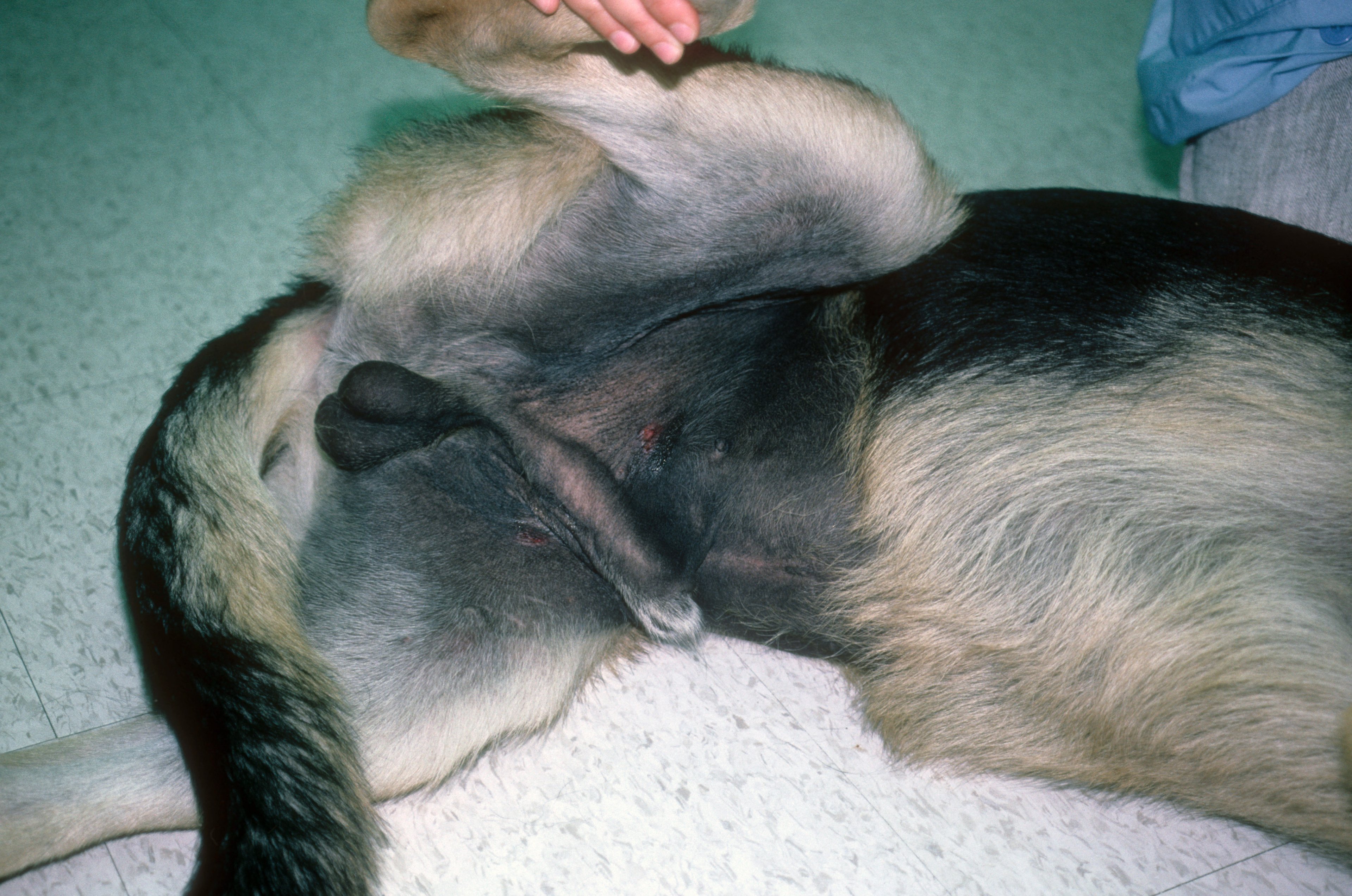
Courtesy of Dr. Karen Moriello.
Postinflammatory hyperpigmentation is triggered by inflammation and/or friction. Lesions can develop into severe areas of hyperpigmentation, with marked lichenification, hair loss, and seborrhea. Often, these areas are odiferous and may be painful. The edges of these lesions are often erythematous; this is a sign of secondary bacterial and/or yeast pyoderma. With time, lesions may spread to the ventral neck, groin, abdomen, perineum, hocks, periocular area, and pinnae. Pruritus is variable and is usually the result of secondary microbial overgrowth (staphylococcal or Malassezia dermatitis) or pruritus from the underlying disease.
Diagnosis of Acanthosis Nigricans
Primary acanthosis nigricans is diagnosed based on physical findings
Postinflammatory hyperpigmentation diagnosis requires identifying the underlying disease
The physical findings compatible with a clinical diagnosis of primary acanthosis nigricans are not difficult to recognize. Postinflammatory hyperpigmentation is a clinical sign of an underlying disease and should be aggressively evaluated through a careful history and physical examination to identify the underlying trigger. Skin scrapings should be performed to exclude demodicosis, and impression smears should be performed to confirm suspected bacterial and Malassezia infections.
Postinflammatory inflammation associated with endocrinopathies is not pruritic, and testing for thyroid and adrenal disease may be useful in older dogs; endocrine skin diseases are not pruritic unless accompanied by secondary skin infections. Intradermal skin testing and/or a food trial may be necessary, but not until parasitic and infectious causes have been ruled out.
Skin biopsies are usually not necessary to confirm primary acanthosis nigricans and are usually not helpful in identification of underlying disease associated with secondary disease. Secondary bacterial and yeast infections are underdiagnosed in this condition, and bacterial culture and skin cytology are important initial tests.
It is now recognized that many cases in Dachshunds are caused by an underlying disease and not a genodermatosis. If a dog is pruritic in the presence of good flea control and infection control, screening for allergic skin disease should be done.
Treatment of Acanthosis Nigricans
Primary acanthosis nigricans may temporarily respond to symptomatic treatment
Postinflammation hyperpigmentation will typically resolve after correction of the underlying cause
Primary acanthosis nigricans in Dachshunds is not curable. In some dogs, lesions do not progress beyond a cosmetic problem. If inflammation is present, early cases may respond to antimicrobial shampoo therapy and local topical glucocorticoids, eg, triamcinolone acetate spray or betamethasone valerate ointment. As lesions progress, more aggressive antimicrobial systemic therapy may be needed but should be based upon culture and susceptibility. Antiseborrheic shampoos are often beneficial for removing excess oil and odor but must be used frequently (ie, 2–3 times/week).
In postinflammatory hyperpigmentation, most of the lesions will resolve after identification and correction of the underlying cause. Some residual lacy hyperpigmentation may remain. Treatment of secondary bacterial and yeast overgrowth is critical. If the dog has not been previously treated for a staphylococcal bacterial infection of the skin, therapy with narrow-spectrum drugs based on culture and susceptibility and/or use of topical chlorhexidine/miconazole shampoos three times a week with chlorhexidine or chlorhexidine/miconazole sprays on non-bath days is recommended.
Culture is recommended to minimize development of methicillin-resistant staphylococci. Yeast infections may be successfully treated with concurrent oral itraconazole or ketoconazole (5 mg/kg). Affected dogs benefit greatly from appropriate antimicrobial therapy and antiseborrheic shampoos (2–3 times/week). If the lesions are caused by friction, emollients may be beneficial.
Clinical signs resolve slowly, possibly over months.
Key Points
What is often diagnosed as acanthosis nigricans is almost always postinflammatory hyperpigmentation and is common in dogs with allergic skin disease and/or frictional apposition.
Pending identification of the underlying skin disease, topical therapy should be used several times a week for relief of pruritus and discomfort and to control odor.
True acanthosis nigricans is a genodermatosis in Dachshunds and is very rare.
For More Information
Also see pet health content regarding hyperpigmentation in dogs.


