


Dermatophytosis (ringworm) is typically a superficial skin infection. It affects a wide range of animals, and several of the causative fungi also cause zoonotic infections. In otherwise healthy animals, it requires no treatment; however, treatment is usually recommended to shorten the course of the disease and avoid contagion. Disease is more common in young or stressed individuals, such as those in extremely crowded environments. Clinical signs can include any combination of hair loss, scaling, crusting, erythema, papules, hyperpigmentation, and variable pruritus. Diagnosis can be confirmed by direct examination of hairs or scales from lesions or by skin biopsy. Dermoscopy or a Wood's lamp can be used to identify hairs for culture and/or direct examination. Fungal culture can determine whether spores are present on the hair coat and must be used in conjunction with clinical examination findings. PCR testing confirms the presence or absence of fungal DNA on the hair coat. It cannot distinguish between viable and nonviable spores. In animals that need treatment, topical antifungal therapy disinfects the hair coat and eliminates infection from hair follicles.
Dermatophytosis is superficial fungal infection of the skin and hair and less commonly of the claw or hoof. The most important pathogens of veterinary importance are:
Microsporum canis (affects cats, dogs, and to a lesser extent large animals)
Trichophyton mentagrophytes, T verrucosum, andT erinacei (affects hedgehogs)
M gypseum (soil organism that causes inflammatory lesions)
The genera Microsporum and Trichophyton are being reclassified into the genus Arthroderma.
Dermatophytosis is a self-curing disease and will resolve without treatment in otherwise healthy animals. It is considered zoonotic, as it causes skin lesions in people that are easily treated. Transmission is by direct contact with an infected animal, but mere exposure does not always result in disease. Transmission from the environment is inefficient if it involves spores alone; microtrauma is needed (eg, clipping of hair coat with contaminated clippers, tack). The prevalence is difficult to determine because it is not a reportable disease in pets, and many studies reporting prevalence do not distinguish between fomite carriage and true infection. Overall, it is an uncommon disease, with reported prevalence of true disease < 4% of all skin disorders. It is not the most common skin disease of cats, contrary to what is reported in lay literature.
Young dogs, free-roaming animals, hunting dogs, and warm environments are risk factors. FIV/FELV-positive status does not predispose cats to infection. Disease is more common in animals under physiologic stress or those in overcrowded environments, such as animal hoarding cases. Infections in animals that are ill, under physiological stress, or have some other underlying disease or factor are more difficult to treat.
Lesion development requires a sufficient amount of infective spores, microtrauma to the skin, and moisture on the skin. The severity of infection reflects the host's overall health; severe infections are not caused by more virulent strains but because of a failure of host cell-mediated immunity to respond to the infection. Dermatophyte arthrospores can germinate and start invading skin and hair shafts within 6–8 hours under ideal conditions.
Clinical Findings
Clinical signs can include any combination of hair loss, scaling, crusting, erythema, papules, hyperpigmentation, and variable pruritus. Nodular lesions (kerion) reactions can develop in dogs. Persian cats can develop nodular lesions (pseudomycetomas). Cats can also develop exudative paronychia. Pustular dermatophytosis can mimic clinical signs of dermatophytosis.

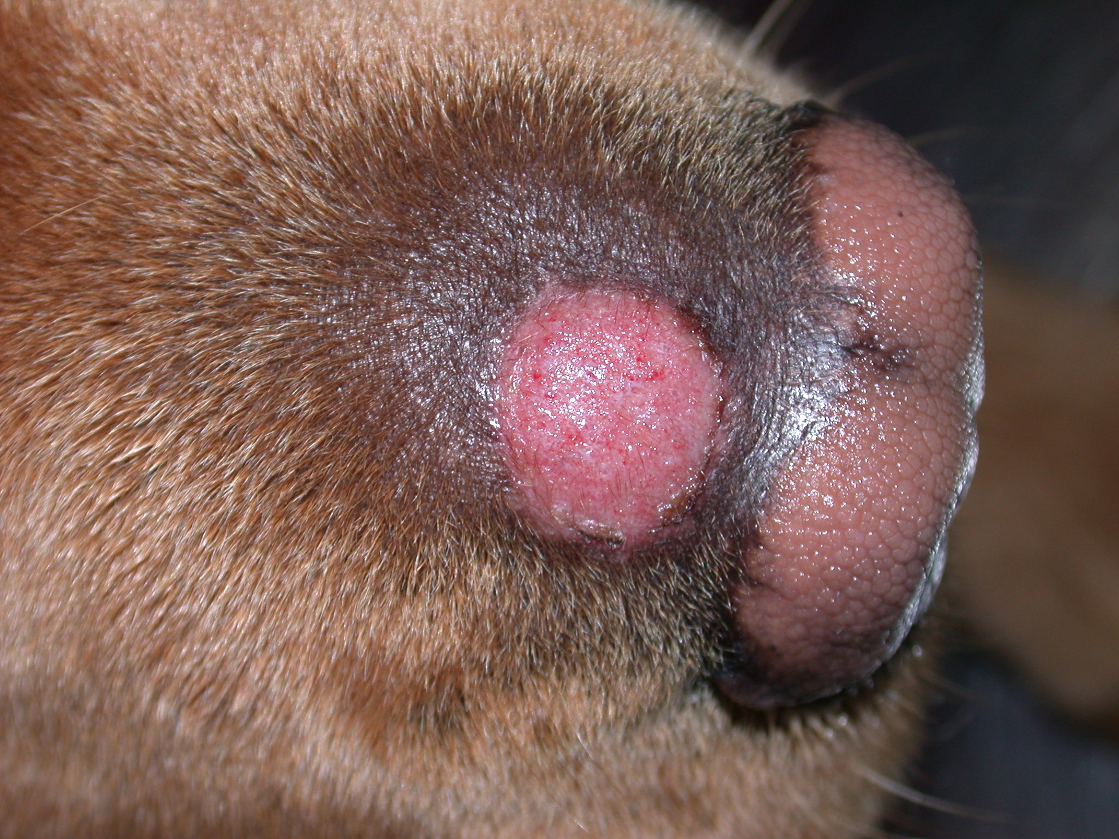
Courtesy of Dr. Sheila Torres.

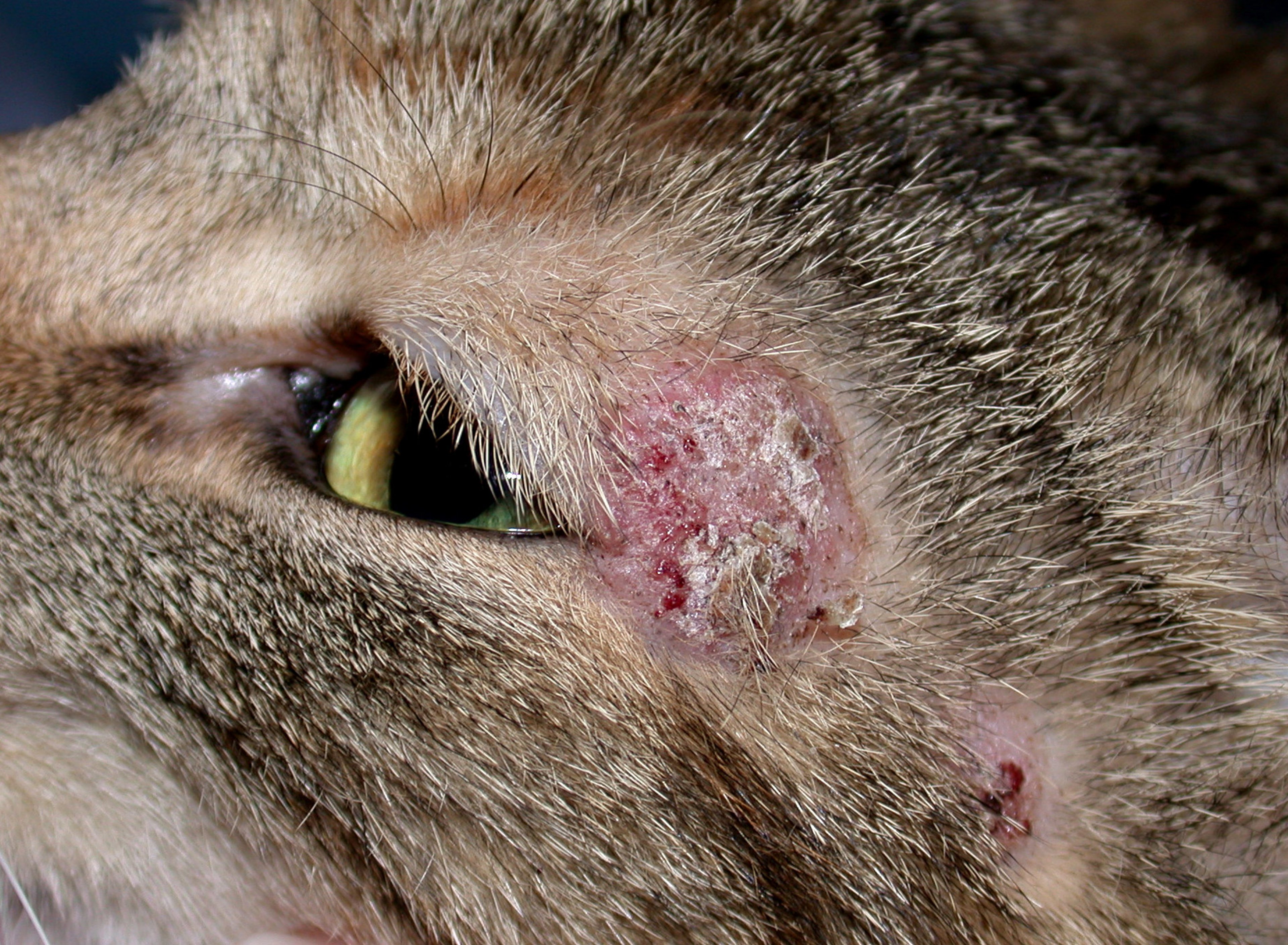
Courtesy of Dr. Sheila Torres.

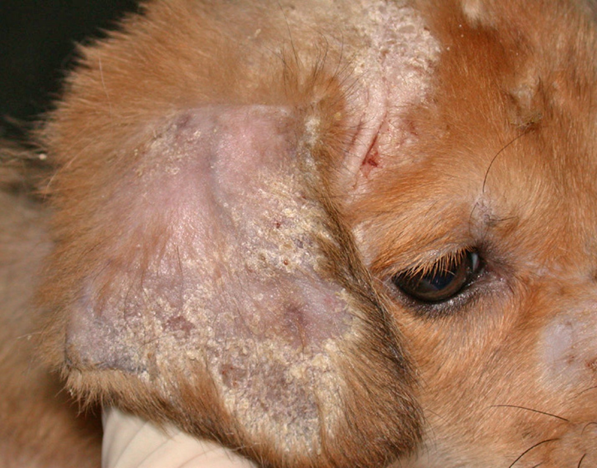
Courtesy of Dr. Sheila Torres.
Diagnosis
Direct examination of scale and hairs or skin biopsy are the only two diagnostic tests that can confirm invasion of the skin/hair follicles.
Bacterial pyoderma in dogs is common and often misdiagnosed as dermatophytosis.
A recent evidence-based review concluded that there is no one test that is the gold standard for diagnosis. Tests are divided into two categories:
Tests that confirm the presence of an active infection in order to make an informed decision (eg, treat or not, euthanize, quarantine)
Tests that confirm the absence of an active infection (eg, the animal poses no infection risk, the animal is cured)
Skin biopsy is not indicated in routine cases of dermatophytosis. It is indicated when there are nodular lesions or an unusual presentation of a skin disease. If it is ordered, the laboratory should be informed this is a possible differential diagnosis so that special stains can be performed. It is important to submit at least several 8 mm skin biopsy punch samples or a nodule. Direct examination of hair and scales scraped and plucked from lesions and mounted in mineral oil can confirm infections in >85% of cases. Clearing agents are not needed.
A Wood's lamp (320 to 400 nm) is a tool used to find M canis-infected hairs. These hairs can be used for direct examination or fungal culture. Positive hairs glow/fluoresce apple green. These hairs are then collected for direct examination via plucking or scraping of material onto a drop of mineral oil. It is important to use a Wood's lamp that is a plug-in with built-in magnification. It does not need to warm up, but the user's eyes must light-adapt to the darkened room. It is important to hold the lamp close to the skin (within 2–4 cm), start at the head, and move slowly. Only hair shafts glow, not crusts. It is important to lift crusts to look for glowing hairs. An evidence-based review found that 91%–100% of untreated, infected animals will have positive glowing hairs. Fluorescence will be less common as animals recover. Sometimes only "glowing tips" will be seen, and these may or may not be culture-positive.
A dermoscope can be used to find abnormal hairs for direct examination. This is a handheld tool used to examine the skin that can locate hairs for direct examination and/or culture.

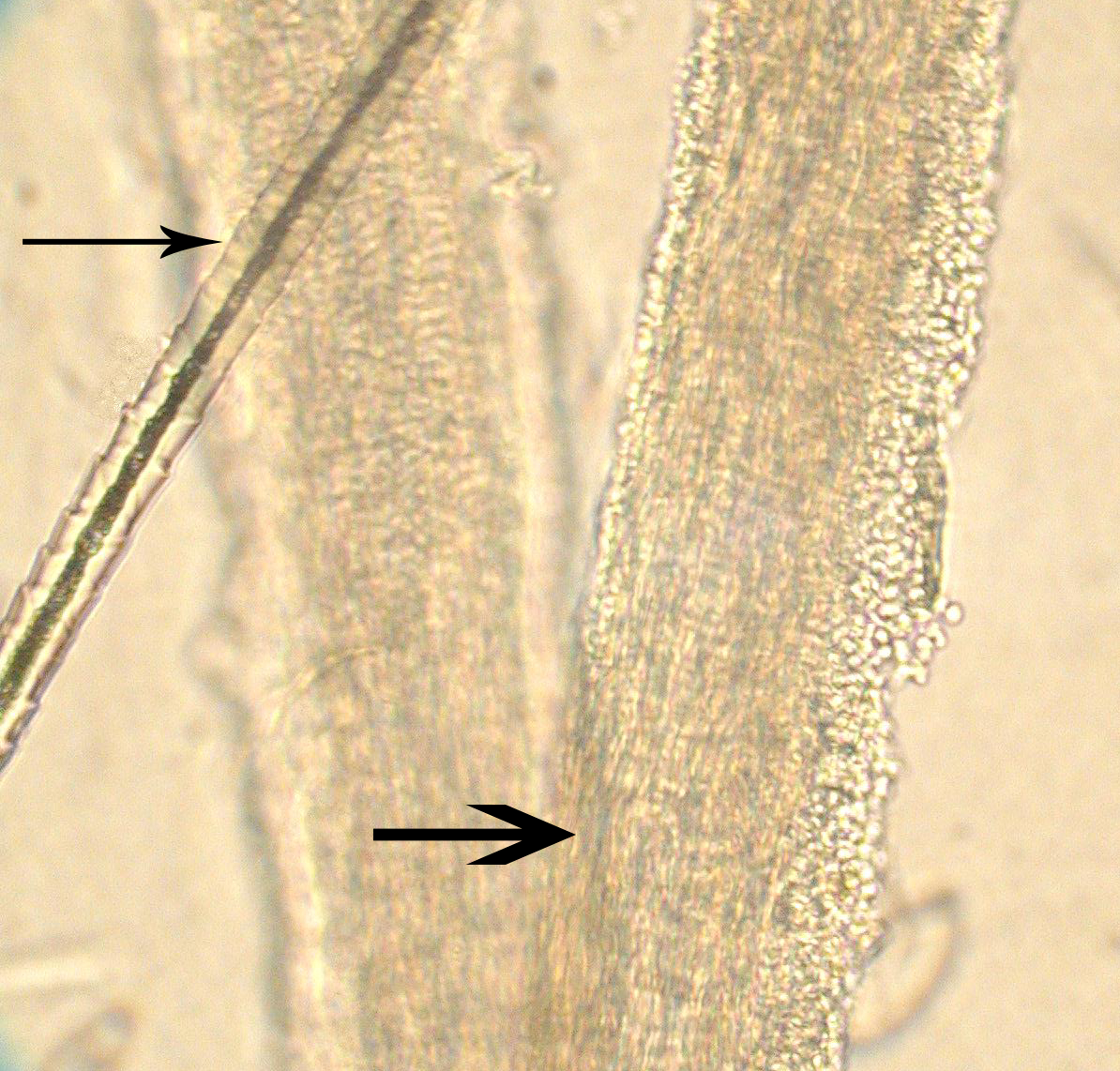
Courtesy of Dr. Karen A. Moriello.
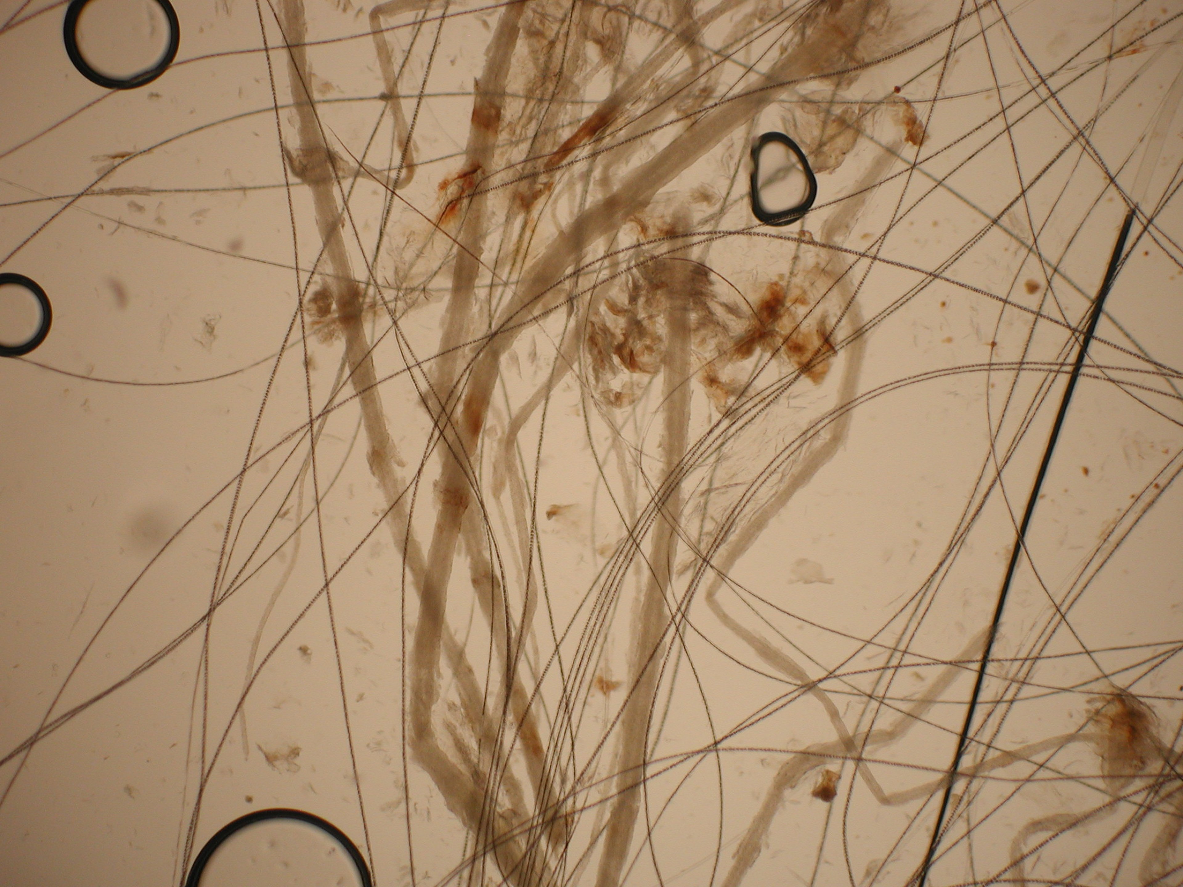
Courtesy of Dr. Karen A. Moriello.
Fungal culture can determine whether spores are present on the hair coat and must be used in conjunction with clinical examination findings. It is best to sample only lesions; the entire hair coat should not be sampled when using this test. For small animals, a new, unopened toothbrush is brushed over the lesions and then inoculated onto fungal culture medium. Plucking hairs from the bristles will increase contamination and should not be done. If plucked hair from lesions is used, it should not be wiped with alcohol, because this may result in a negative culture. Fungal cultures can be done as a point of care test only if the user follows directions for storage and use and does daily gross and microscopic examination of colonies. If this is not possible, a diagnostic laboratory should be used. M canis and M gypseum are not difficult to isolate and identify. Trichophyton species are hard to identify to genus level.
Fungal culture key points:
Use flat plates that are easy to inoculate. Do not use jars or small flat plates intended for samples from people.
Stab bristles of toothbrush onto the surface 5 to 6 times; do not over-inoculate the plate or sporulation will not be seen.
Incubate at room temperature; seal the plate in a plastic bag to prevent desiccation.
Examine daily for 14 days.
Suspect pathogens are pale or light in color and never heavily pigmented.
If dermatophyte test medium is used there will be a color change (red) around the colony.
Post-treatment cultures can have abnormal gross and microscopic appearance.
Pathogen type can be confirmed by direct examination of the colony (adhesive tape prep) using lactophenol cotton blue stain.
PCR testing confirms the presence or absence of fungal DNA on the hair coat. It cannot distinguish between viable and nonviable spores. Availability of the validated testing protocols limits use. It is important to submit adequate samples to the laboratory (>20 hairs and crusts).
Treatment
Dermatophytosis is a self-curing disease in most animals
In animals that are treated, a systemic antifungal drug will eliminate active infection in hair follicles
Animals can be treated to shorten the course of the disease and minimize contagion to other susceptible animals or people. Infected small animals should be kept isolated from other pets until there is clear evidence of clinical cure. Young animals should not be overly confined or undersocialized, or lifelong behavioral problems may occur.
Cats can be treated with itraconazole (5 mg/kg, PO, once daily on a week on/week off schedule). Most infections are resolved after 3 or 4 cycles. An evidence-based review found that itraconazole is well tolerated in cats and was not associated with liver toxicity or vasculitis. Compounded itraconazole should not be used however. Poor bioavailability has been documented for such formulations, so a commercial veterinary liquid formulation should be used.
Small dogs can be treated with oral itraconazole (5 mg/kg, once daily); pulse therapy is likely to be effective, but this has not been documented. Other dogs can be treated with ketoconazole (5 mg/kg, once daily) or terbinafine (30–40 mg/kg, once daily).
Other points to keep in mind regarding drug therapy for dermatophytosis include:
Ketoconazole should not be used in cats because it causes anorexia.
Fluconazole should not be used because this is the least effective drug for dermatophytes.
Griseofulvin is no longer recommended because itraconazole and terbinafine are superior drugs.
Lufenuron is ineffective.
In addition to systemic treatment, topical therapy is required because it disinfects the hair coat. This is important because infective spores are the source of contagion and transmission, and disinfection minimizes environmental contamination. A whole body rinse (lime sulfur 1:16 or enilconazole 1:100) should be used twice a week; these products have residual activity. Shampoo containing 2% chlorhexidine and 2% miconazole is effective and may be the only option in countries where lime sulfur or enilconazole is not available; shampoo therapy has no residual activity. Even if systemic therapy is stopped, topical therapy should be continued until a mycologic cure is demonstrated.
Adjunct focal topical therapy can be used for lesions in hard-to-treat locations such as the ears and face. A 1%–2% vaginal miconazole cream can safely be used on the face. For the ears, otic products that contain clotrimazole or miconazole/chlorhexidine or ketoconazole/chlorhexidine combinations are available.
Environmental cleaning removes infective material from the environment. Spores do not multiply in the environment and do not invade the environment like mildew. Spores are a normal dormant life stage of dermatophytes and other microorganisms and are easily removed from the environment. Mechanical removal of organic material and hair followed by washing of the surface with a detergent until visibly clear is the most important step for environmental cleaning/disinfection. After cleaning, a disinfectant should be used. Any bathroom disinfectant labelled as effective against Trichophyton will kill any spores not removed by mechanical cleaning. Disinfectant use alone will not remove environmental contamination. Thorough cleaning once or twice a week is adequate. Between cleanings, remove organic material with wipes or other means. Soft surfaces such as bedding or towels can be disinfected via washing; bleach is not needed. Carpets can be disinfected via carpet shampooing or steam cleaning.
The end point of treatment includes both clinical and mycologic cure. Clinical cure is the resolution of all lesions and the lack of any new lesions. A Wood's lamp examination can be used to look for areas of residually infected hairs in animals with M canis infections. Once lesions have resolved and there is a clear clinical cure, a fungal culture or PCR can test for mycologic cure. One negative PCR test supports a mycologic cure. Unless the animal has systemic illness, one negative fungal culture also supports mycologic cure.
Key Points
Dermatophytosis is caused by several Microsporum and Trichophyton spp and can affect a wide range of species.
Infections in otherwise healthy animals are self-limiting and do not require treatment.
Animals can be treated to shorten the course of the disease and minimize contagion to other susceptible animals or people.
In animals that are treated, a systemic antifungal drug will eliminate active infection in hair follicles.
In addition to systemic treatment, topical therapy is required because it disinfects the hair coat.
For More Information
Also see Pet Health content regarding ringworm in dogs and ringworm in cats.


