


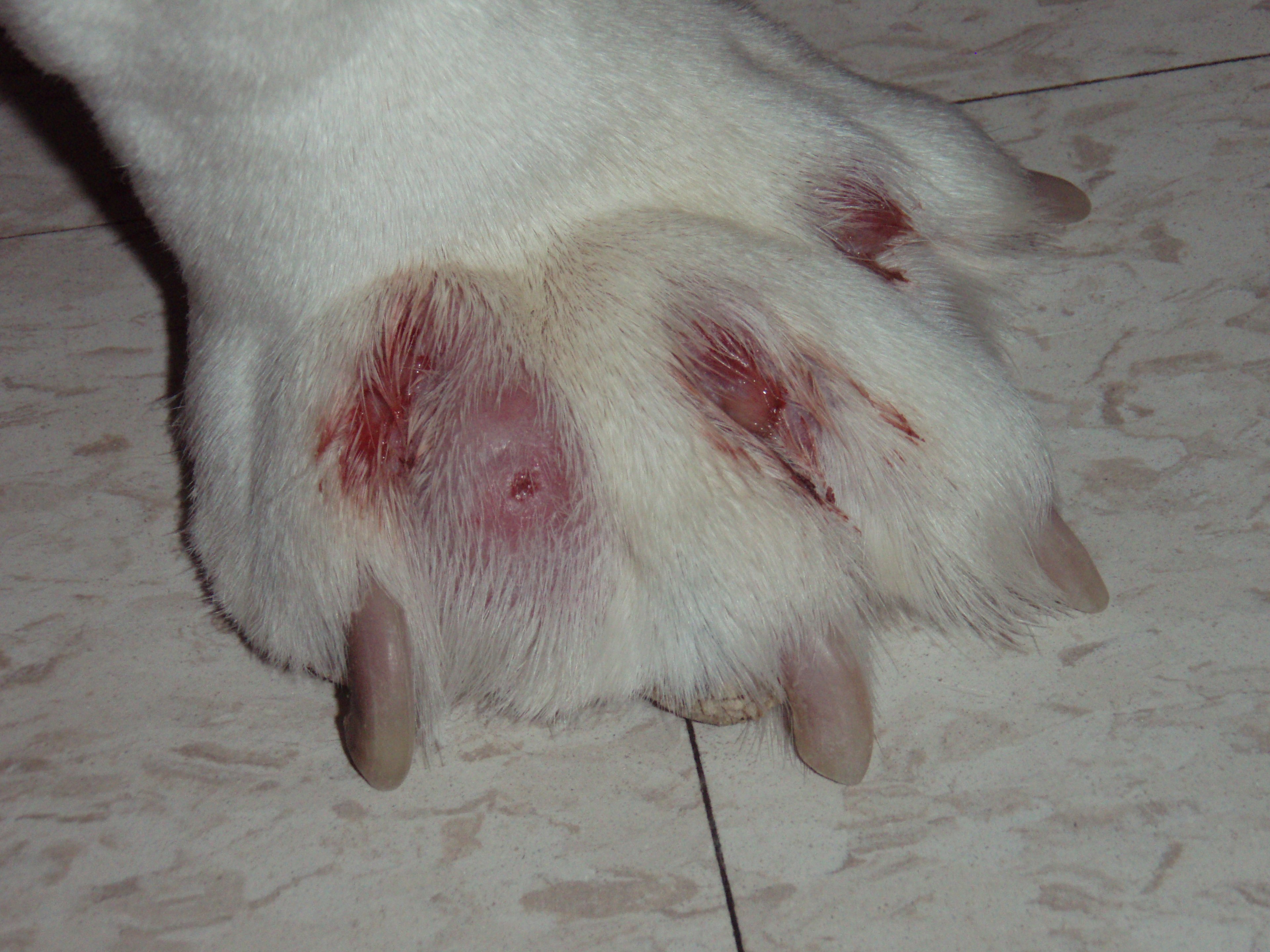
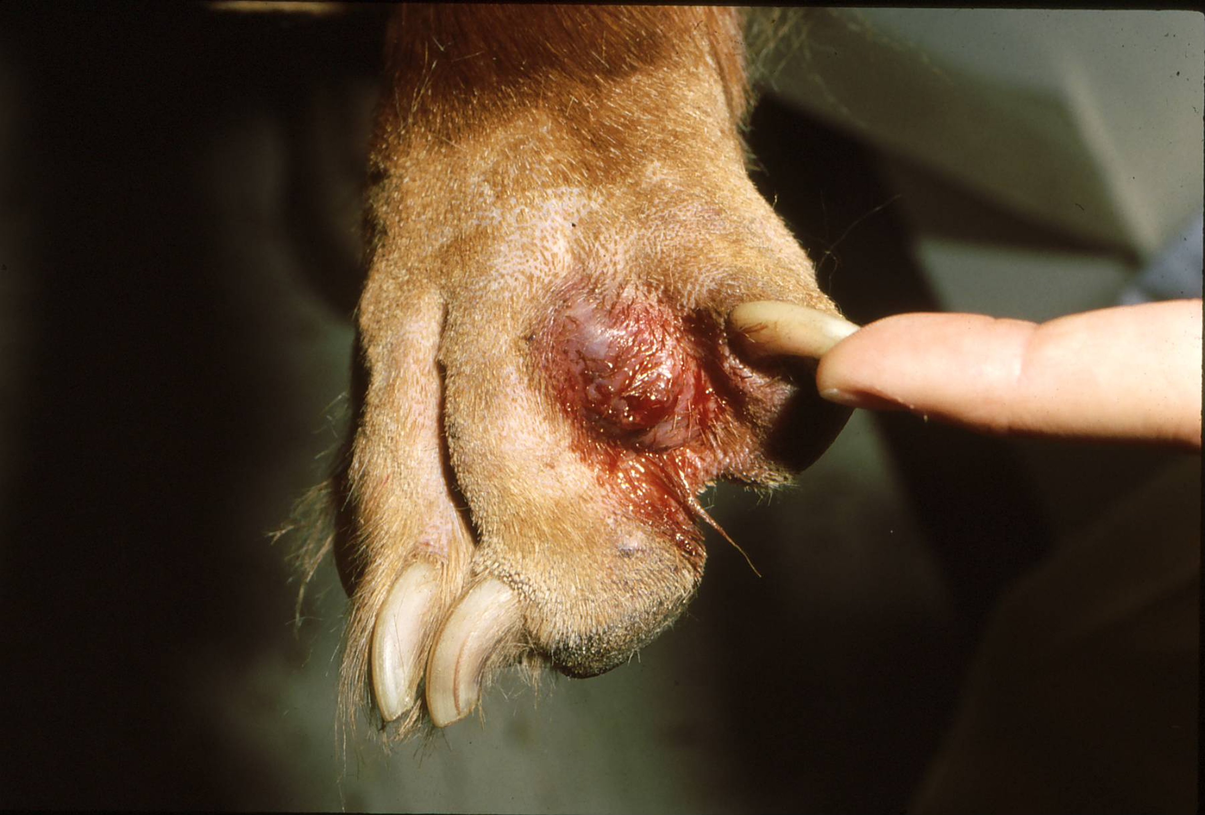
Interdigital furuncles are deep pyoderma lesions that form between the toes of dogs. They can be either single or multifocal. The nodules are painful areas of pyogranulomatous inflammation. Foreign body reactions to embedded hair shafts will prolong the infection. Lesions are treated with topical antimicrobial therapy and, if severe, with concurrent systemic antibiotics. It is important to identify the underlying trigger (eg, atopic dermatitis) if lesions are multifocal.

Courtesy of Dr. Karen A. Moriello.

Courtesy of Dr. Karen A. Moriello.
Interdigital furuncles are areas of deep pyoderma. Interdigital furuncles are often incorrectly referred to as interdigital cysts. Clinically, these lesions are painful, focal or multifocal nodules in the interdigital webs of dogs. Histologically, these lesions represent areas of nodular pyogranulomatous inflammation—they are almost never cystic. Canine interdigital palmar and plantar comedones and follicular cysts is a recognized syndrome that may be a subtype of interdigital furuncles or a separate disease. Foreign body reaction to embedded keratin hair shafts is a major obstacle to resolution of infection.
Etiology of Interdigital Furunculosis in Dogs
The most common cause of interdigital furunculosis is a deep bacterial infection. Many dog breeds (eg, Chinese Shar Pei, Labrador Retriever, English Bulldog) are predisposed to bacterial interdigital furunculosis because of the short bristly hairs located on the webbing between the toes, prominent interdigital webbing, or both. The short shafts of hairs are easily forced backward into the hair follicles during locomotion (traumatic implantation). Hair, ie, keratin, is very inflammatory in the skin, and secondary bacterial infections are common. Less commonly, foreign material is traumatically embedded in the skin. Demodicosis may be a primary cause of interdigital furunculosis. Canine atopic dermatitis is also a common cause of recurrent interdigital furunculosis.
The cause of canine interdigital palmar and plantar comedones and follicular cysts is unknown but most likely involves trauma, resulting in epidermal and follicular infundibular hyperkeratosis, acanthosis, plugging or narrowing of the follicular opening, and retention of the follicular contents.
Clinical Findings and Lesions of Interdigital Furunculosis in Dogs
Early lesions of interdigital furunculosis may appear as focal or generalized areas of erythema and papules in the webbing of the feet that, if left untreated, rapidly develop into single or multiple nodules. The latter usually are 1–2 cm in diameter, reddish purple, shiny, and fluctuant; they may rupture when palpated and exude a bloody material. Interdigital furuncles are most commonly found on the dorsal aspect of the paw but may also be found ventrally. Furuncles are usually painful, and the dog may be obviously lame on the affected foot (or feet) and lick and bite at the lesions. Lesions caused by a foreign body, eg, a grass awn, are usually solitary and are often found on a front foot; recurrence is not common in these cases. If bacteria cause the interdigital furunculosis, there may be several nodules with new lesions developing as others resolve. A common cause of recurrence is the granulomatous reaction to the presence of free keratin in the tissues.
Dogs with interdigital comedones and follicular cysts typically present with lameness and draining tracts. Skin lesions are not often seen unless the hair coat is clipped. Areas of alopecia and thickened, firm, callus-like skin with multiple comedones are typical.
Diagnosis of Interdigital Furunculosis in Dogs
Typically based on clinical signs
For furunculosis, the diagnosis is often based on clinical signs alone . The major differential diagnoses are traumatic lesions, foreign bodies, follicular comedone cysts, and neoplasia, although the latter is rare. The most useful diagnostic tests include hair trichograms for Demodex mites, impression smears, and fine-needle aspirates to confirm the presence of an inflammatory infiltrate. A commonly overlooked concurrent infection is Malassezia. Bacterial culture and susceptibility is recommended to guide antimicrobial use. Unusual or recurrent lesions should be excised for histopathologic examination. Solitary lesions may require surgical exploration to find and remove foreign bodies such as grass awns.
Definitive diagnosis of palmar and plantar follicular cysts requires a skin biopsy. However, these cysts are suspected when clinical examination reveals draining tracts associated with callus-like lesions or obvious comedone formation. Moderate to extensive compact hyperkeratosis and acanthosis of the epidermal and follicular infundibulum is found. Follicular cysts consisting of keratin are common. Often, lesions are complicated by secondary infection and concurrent bacterial furunculosis.
Treatment of Interdigital Furunculosis in Dogs
Topical antibiotics are always indicated, and severe cases may require systemic therapy
Topical therapy is always indicated in cases of interdigital furunculosis. Severe cases may need concurrent systemic antibiotic therapy. Treatment is best based on culture and susceptibility, because these are deep infections and may require longterm therapy, particularly if multifocal.
Do not clip the paws with electric clipper blades, because this may cause microtrauma and lead to hair shafts becoming traumatically inoculated into tissue. Use scissors to clip hair.
Pending culture, instruct clients to wash paws daily with a combination of 2% chlorhexidine/2% miconazole shampoo.
Pending culture, apply a polymyxin B and bacitracin ointment several times a day.
If there is a concurrent Malassezia overgrowth, administer ketoconazole 5 mg/kg once daily or, in small dogs, itraconazole 5 mg/kg once daily.
Administer systemic antibiotic therapy based upon culture and susceptibility findings for no less than 4 weeks.
Dogs with multidrug-resistant methicillin resistant staphyloccocal infections may benefit from topical mupirocin ointment.
Some dogs have chronic recurrent lesions despite good initial treatment and identification of an underlying disease. These dogs are best treated with chronic topical antimicrobial bathing.
Chronic, recurrent interdigital furunculosis is most often caused by inappropriate antibiotic therapy (too short a course, wrong dosage, wrong drug), concurrent systemic corticosteroid administration, demodicosis, an anatomic predisposition, or a foreign body reaction to keratin. Lesions that recur despite therapy can also be a sign of an underlying disease, eg, atopy, hypothyroidism, or concurrent Malassezia infection. Lesions in confined dogs are likely to recur unless the dog is removed from wire or concrete surfaces.
Treatment of interdigital palmar and plantar comedones and follicular cysts can be successfully accomplished by laser therapy. Postoperative care is time intensive, with hydrotherapy and bandage changes once or twice daily.
Key Points
Interdigital bacterial furuncles are solitary or multiple areas of deep pyoderma that may be triggered by trauma, conformation, or underlying skin disease. Chronic lesions are often caused by foreign body reactions to embedded keratin.
Topical therapy (bathing and overall hygiene) is a key part of initial therapy and for chronic, recurrent lesions. Combined chlorhexidine/miconazole shampoo products are recommended.
Systemic antibiotic therapy and concurrent topical therapy are often needed when lesions are multiple and painful.
For More Information
Also see pet health content regarding abscesses between the toes in dogs.


