


Courtesy of Dr. Ronald Green.
Elbow dysplasia is a generalized incongruency of the elbow joint in young, large, rapidly growing dogs that is related to abnormal bone growth, joint stresses, or cartilage development. One or more of the following lesions may be present in the joint: an ununited anconeal process of the ulna, fragmentation of the medial coronoid process of the ulna, and osteochondrosis of the medial aspect of the humeral condyle. Radiographic grading of dysplastic elbow joints is performed by the Orthopedic Foundation for Animals in the USA and in Scandinavian and European kennel clubs.
Ununited Anconeal Process in Elbow Dysplasia in Dogs
This results when there is separation of the ossification center of the anconeal process from the proximal ulnar metaphysis. Fusion should be completed by 5–6 months of age. The fracture is postulated to result from a biomechanical imbalance of force and movement in the rapidly growing elbow. Initially, the anconeal process is connected to the ulna by a bridge of fibrous tissue, which fragments to form a pseudoarthrosis, and the elbow becomes unstable. This joint laxity continues to damage the articular cartilage, and secondary osteoarthritis results. A hereditary basis has been implicated but not proved.

Courtesy of Dr. Joseph Harari.
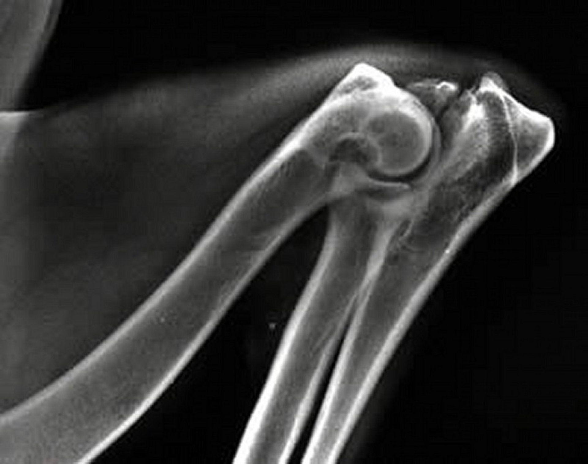
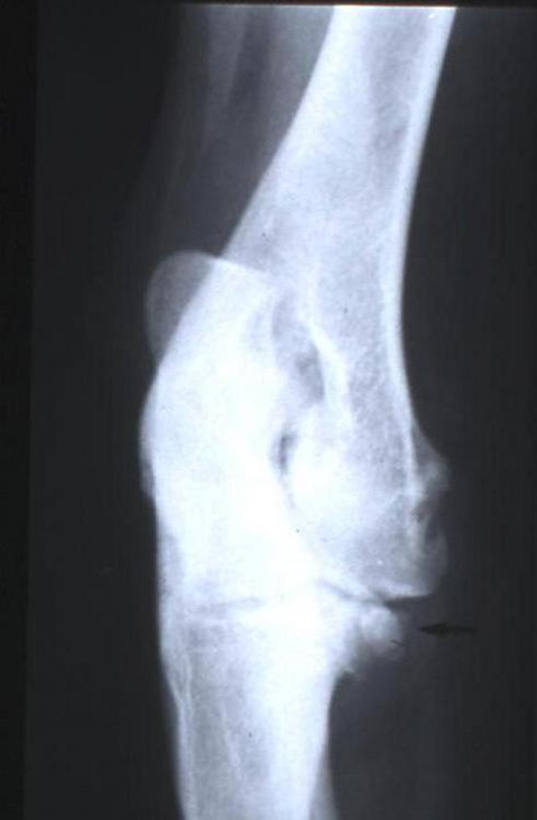
Lameness develops insidiously between 4 and 8 months of age; however, some bilateral cases may not be diagnosed until dogs are >1 year old. Affected elbows may deviate laterally, and the range of motion is restricted. Advanced cases have osteoarthritis, joint effusion, and crepitus. Clinical signs are suggestive, and the diagnosis is confirmed by radiography. A lateral radiograph of the elbow in the flexed position allows visualization of the ununited process. Both elbows should be examined because the condition can be bilateral.
Fragmentation of the Medial Coronoid Process in Elbow Dysplasia in Dogs
In this condition of the medial compartment of the canine elbow, the coronoid process fails to unite, either partially or totally, with the ulnar diaphysis and, thus, does not become a part of the articular surface of the trochlear notch. Joint laxity, irritation, and finally osteoarthritis result. This condition and osteochondrosis of the medial humeral condyle are considered to be the most common causes of osteoarthritis of the canine elbow. Bone fragments can be seen by radiography, arthroscopy, or CT.
Osteochondrosis of the Medial Humeral Condyle in Elbow Dysplasia in Dogs
This results from a disturbed endochondral fusion of the epiphysis of the medial epicondyle with the distal end of the humerus. The exact cause is unknown, but because the carpal and digital flexors originate from the ventral aspect of this structure, it may represent an epiphyseal avulsion. It results in pain on flexion of the elbow or deep digital palpation and is accompanied by soft-tissue swelling. Radiographically, radiodense structures have been seen caudal and distal to the area of the medial epicondyle.
Treatment of Elbow Dysplasia in Dogs
Early surgical treatment is recommended before degenerative joint disease develops. For fragmentation of the medial coronoid process, a medial arthrotomy or arthroscopy is performed and the fragmented process removed. For ununited anconeal process, either a lateral arthrotomy is performed and the ununited process removed, or a midshaft ulnar osteotomy is performed to relieve asynchronous growth and result in union of the process. Reattachment of the process by screw fixation is also an option. For osteochondrosis, the subchondral bone lesion is curetted to stimulate fibrocartilage formation. Transplantation of cartilage plugs in lieu of curettage has recently been described. Prognosis after surgery is good if degenerative joint disease has not developed in the joint. Aspirin or NSAIDs (eg, carprofen, deracoxib, firocoxib, meloxicam, tepoxalin) can be used to reduce pain and inflammation. Joint-fluid modifiers (glycosaminoglycans, hyaluronic aid) and acupuncture may be useful, although most reports are anecdotal. Joint injection with mesenchymal cells or platelet-rich plasma has also been described; reports are inconclusive.
For More Information
Also see pet health content regarding elbow dysplasia in dogs and joint disorders in cats.


